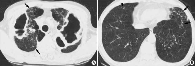
Fig. 1.
A 66-yr-old man with M. avium-intracellulare complex pulmonary disease. (A) Transaxial thin-section (2.5-mm thickness) CT scan obtained at level of great vessels shows multiple large thin-walled cavities in both lung apices. Also note several small nodules (arrows) in right lung. (B) CT (2.5-mm thickness) scan obtained at level of suprahepatic inferior vena cava shows multiple small nodules and branching centrilobular nodules, so-called tree-in-bud pattern (arrows), in lingular segment of left upper lobe and left lower lobe. Small nodule (arrowhead) is also seen in right middle lobe.

