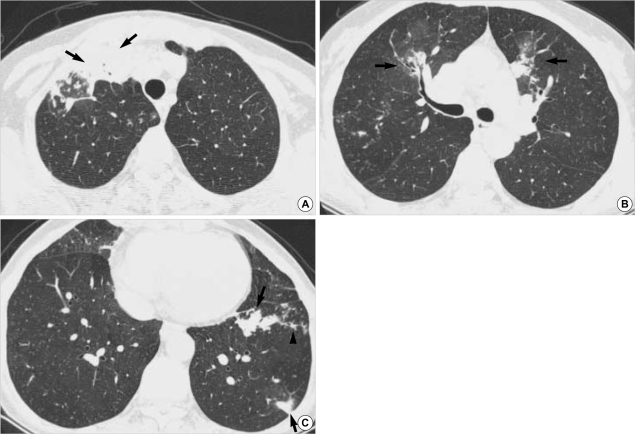
Fig. 3.
A 52-yr-old woman with M. avium-intracellulare complex pulmonary infection. (A) Transaxial thin-section (2.5-mm thickness) CT scan obtained at level of great vessels shows subsegmental consolidation with open bronchus sign in right upper lobe (arrows). (B) CT (2.5-mm thickness) scan obtained at level of right upper lobar bronchus shows airspace consolidation with surrounding ground-glass opacity (arrows) along bronchovascular bundles in anterior segments of both upper lobes. Also note small nodules in right upper lobe. (C), CT (2.5-mm thickness) scan obtained at lung base shows lobular consolidation in left lower lobe (arrows). Also note lesion of tree-in-bud pattern (arrowhead) in left lower lobe and small nodules in right middle lobe.

