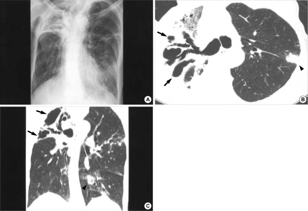
Fig. 1.
M. intracellulare pulmonary disease of the upper lobe cavitary form in a 73-yr-old man. Chest radiograph shows parenchymal opacity which is containing multiple cavities in the right upper lobe. Also note nodular lesions in left middle and lower lung zones (A). Transaxial lung window CT image (2.5-mm section thickness, 70 mA) obtained at the level of the right upper lobar bronchus shows dilated bronchi in an opacified right upper lobe. Also note associated multiple cavitary lesions (arrows). A lobular consolidative lesion (arrowhead) is seen in the left upper lobe (B). Coronal reformation (2.0-mm section thickness) image demonstrates dilated bronchi and multiple thin walled cavities (arrows) in the right upper lobe. Also note multiple variable-sized nodules and consolidation (arrowhead) in the left lung (C).

