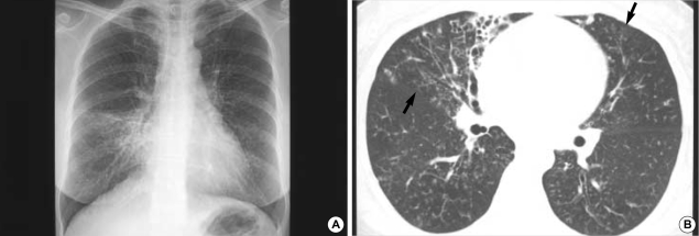
Fig. 3.
M. abscessus pulmonary disease in a 49-yr-old woman. Chest radiograph shows multifocal patchy areas of small nodular clusters in both lungs. Also note parenchymal opacity in the right middle lobe (A). Transaxial lung window CT image (2.5-mm section thickness, 70 mA) obtained at the basal trunk (B) show bronchiectasis and small centrilobular nodules or tree-in-bud opacities (arrows) in the right middle lobe and in the lingular division of the left upper lobe. Also note bronchiolitis of small centrilobular nodules and tree-in-bud opacities in both lower lobes.

