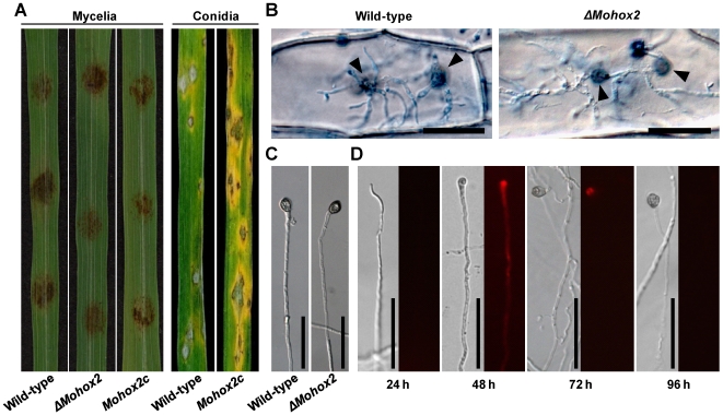
Figure 3. Effect of MoHOX2 deletion on appressorium development and pathogenicity.
(A) Assay for pathogenicity. Intact rice leaves were inoculated either by placement of agar plugs containing mycelia (6 mm in diameter) or by spraying conidial suspension (105 conidia/ml) of the indicated strains. (B) Assay for hypha-mediated penetration. Onion epidermis was inoculated with a patch of mycelia of the wild-type or ΔMohox2. Black arrowheads indicate sites of penetration via hyphal appressoria. Bars = 50 µm. (C) Appressorium development at hyphal tips. Mycelial blocks were placed on hydrophobic surfaces, and incubated for 72 h at 25°C. Bars = 50 µm. (D) Microscopic observation of temporal and spatial occurrence of lipid droplets during hypha-driven appressorium development. Mycelial blocks of the wild-type were incubated on hydrophobic surfaces, and stained with Nile red to visualize the formation of lipid droplets. Note that lipid droplets were not initially detected in hypha until 24 h, but became abundant in hypha with developing apprssorium 48 h after inoculation. Bars = 50 µm.

