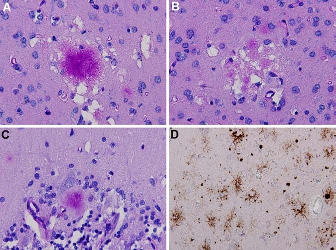
Fig. 2.

Neuropathology of variant CJD: A. florid plaque surrounded by small areas of spongiform change in the occipital cortex (Luxol-PAS stain). B. Multiple small cluster plaques in the occipital cortex (Luxol-PAS stain). C. kuru-type plaque in the cerebellar cortex (Luxol-PAS stain). D. Immunohistochemistry for prion protein (using monoclonal antibody 3F4) shows strong staining of plaques in the occipital cortex, along with amorphous and pericellular deposits
