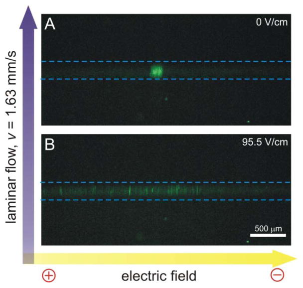
Figure 2.
Dispersion of mitochondria in μFFE caused by application of an electric field. The mitochondria stream is narrow in the absence of an electric field (A) and disperses when a 95.5 V/cm field is applied (B). Images were produced as an overlap of 60 subsequently captured images (scan rate 10 Hz). Dashed lines were added to illustrate the detection region illuminated by the Ar+ laser. Bright spots outside the detection zone were caused by the light scattered at irregularities in the microchip formed during the fabrication process. μFFE buffer: 250 mM sucrose, 10 mM HEPES, pH=7.4; flow rate: 500 μL/min; Sample flow rate: 250 nL/min; LIF detection: 488 nM excitation, fluorescence collection using 2× magnification, 525±25 band pass filter, CCD camera integration: 100 ms.

