Abstract
In vivo, mammalian lenses have the capacity to effect fully reversible changes in shape, and possibly volume, during the accommodation process. Isolated lenses also change shape by readily swelling or shrinking when placed in anisotonic media. However, the manner by which the lens changes its shape when its volume is changed osmotically is not firmly established. Putatively, the lens could swell or shrink evenly in all directions, or manifest distinctive swelling and/or shrinking patterns when exposed to anisotonic media. The present study measured physical changes in lenses consistent with the latter alternative using methods we developed for determining rapid changes in lens shape and volume. It was found in isolated rabbit and cow lenses that the length of the axis between the anterior and posterior poles (A-P length) primarily increases under hypotonic conditions (−40 to −100 mOsM), with smaller, or no changes, in equatorial diameter (ED). Hypertonic conditions (+50 to +100 mOsM) on rabbit lenses elicited a predominant reduction in ED, while the A-P length was only marginally reduced. Hypertonic solutions of +150 mOsM were required to obtain similar changes in cow lens shape. The ratio of the A-P length to the ED was taken as a measure of “circularity”. This ratio increased gradually in rabbit and cow lenses bathed in hypotonic solutions because of the increase in the A-P length. The calculated lens volume increased in tandem with the increase in “circularity”. Lens circularity also increased under hypertonic conditions due to the decrease in ED, but this increase in circularity during shrinkage was not as pronounced as that which occurred during swelling. As such, the lens has a tendency upon swelling to change its shape by approaching the structure of a globular spheroid (as occurs during accommodation for near focusing), but lens shrinkage does not result in a flatter lens with a reduced A-P length as occurs during dis-accommodation for distance focusing. Moreover, osmotically evoked shape changes appear irreversible, in contrast to the mechanically elicited shape changes of accommodation.
Keywords: lens shape, lens volume, lens swelling, lens shrinkage, lens osmometer, lens volume regulation
Introduction
The crystalline lens is a highly well-ordered physical structure (Kuszak, 1995; Zampighi et al., 2002; Kuszak et al., 2006) with a morphology conducive to its remarkable transparency and its ability to change shape during the accommodation process. It gets rounder for closer vision and flatter for distant vision, but its most remote shape remains exactly the same in each of the of the two extremes. In vivo, human lenses may accommodate over the course of decades millions of times, but the process always involves a completely reversible change in shape, and possibly volume (Gerometta et al., 2007). In the present work, we aimed to determine if a range of anisotonic media could simulate lens shape changes in accommodation.
We recently showed with isolated lenses under conditions mimicking accommodation that changes in lens volume could be calculated in accord with the shape changes (Gerometta et al., 2007; Zamudio et al., 2008). In these works, we described in detail our method to calculate volume from lens cross-sectional area (CSA). Presently, this approach was used to monitor the manner by which the lens changes its shape when its volume is altered under anisotonic conditions.
Hypothetically, the lens could change its shape uniformly when exposed to perturbations in bath osmolality (i.e., swell or shrink evenly in all directions), or asymmetrically (i.e., the shape changes may occur in a manner analogous to the asymmetric shape changes exhibited by the lens during the accommodation process). To the best of our knowledge, only one study considered this aspect and reported that bovine lenses swell asymmetrically in a hypotonic medium. Zhang and Jacob (1996) found that only the length of the axis between the anterior and posterior poles (A-P length) increased and that the equatorial diameter (ED) was unaffected. We have corroborated this observation. In addition, we also examined the effects of hypertonic conditions on lens shape to see if lens shrinkage results in a decrease in A-P length as occurs during dis-accommodation when the lens becomes flatter. Instead we observed in both rabbit and cow lenses a reduction in ED, without a marked effect on A-P length. We examined the effects of anisotonic media on lens shape and volume in two different species to see if the evoked changes were common phenomena.
Zhang and Jacob (1996), as well as Patterson and Fournier (1976), reported calculated lens volumes for bovine and rat lenses bathed in hypotonic media but used a relatively crude calculation by applying the equation for the volume of a sphere to the lens, an approach that may be somewhat more accurate for the more spherical rat lens, but not for lenses of cows and rabbits. Moreover, other classical studies characterizing lenticular hydration state under anisotonic conditions recorded changes in lens wet weight and/or water content as a function of time of exposure to the altered milieu (commonly for a period of hours), but did not measure changes in lens shape or volume (Kinoshita et al., 1965; Cotlier et al., 1968).
Finally, we obtained within a 10–20 min time frame, linear changes in rabbit lens volume over a −90 to +100 mOsM tonicity range, a response that apparently occurred in the presence of a simultaneous volume regulation by the whole lens as judged by the limited extent of the volume changes to the imposed anisotonic challenge. However, the tonicity shifts used were too large to obtain volume and shape recoveries within the time frame of the present experiments (even when the control conditions were restored). This underlines the small range under which lens shape can be reversibly changed. It appears possible that reversible volume changes may be obtained with shorter exposure to the external forces.
Materials and Methods
Lens Bathing Solutions
An isotonic modified Tyrode’s solution was used to nourish the lens at the beginning of experiments. It had the following composition (in mM): 1.8 calcium gluconate, 1.2 MgCl2, 4.5 KCl, 105 NaCl, 30 NaHCO3, 1 NaH2PO4, 10 glucose, and 15 sucrose. The pH of this solution when bubbled with 5% CO2-95% air was 7.5. It measured 290 mOsmole/kg water.
In this work, the hypertonic solutions are referred to as the difference between isotonicity (i.e., 290 mOsM) and the calculated value for the hypertonic osmolality with a positive sign. For hyposmotic solutions, the difference between isotonicity and the calculated hypotonic osmolality are indicated with a negative sign.
For experiments with rabbit lenses, hypertonic media (+50 and +100 mOsM) were prepared by adding appropriate amounts of sucrose into the above Tyrode’s solution. With cow lenses, hypertonic solutions of +100 and +150 mOsM were used.
Hypotonic solutions for the rabbit lens experiments (−40 and −90 mOsM) were obtained by diluting the above control Tyrode’s with suitable portions of a solution containing 5 mM Trizma base, 2.5 mM KCl, 1.0 mM CaCl2 and 1.0 mM MgCl2. For the cow lens experiments, hypotonic solutions of −100 mOsM were prepared.
Dissection and Mounting of the Rabbit and Cow Iris-Ciliary Body-Zonulae-Lens Complex
Experiments using rabbit lenses were done in New York and those using cow lenses were done in Corrientes, Argentina. In the former case, adult albino rabbits of either sex weighing 3–4 kg were purchased commercially and rapidly killed by CO2 asphyxiation, a protocol approved by the Mount Sinai Hospital Animal Care and Use Committee. Prior to sacrifice, the rabbits’ care conformed to the ARVO Statement for the Use on Animals in Ophthalmic and Vision Research. Experiments using cow eyes were done in the laboratory of Dr. Gerometta at the School of Medicine of UNNE. The cow eyes were obtained from a nearby slaughterhouse and transported on ice to UNNE within 0.5 hr post-mortem.
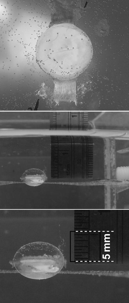
The enucleated rabbit and cow eyes were dissected as described previously by Gerometta et al. (2007) to obtain the iris-ciliary body-zonulae-lens complex (referred to below as the ICB-lens complex). For this, most of the cornea was removed via a circular cut 1–2 mm anterior to the limbal region. The attachment between the sclera and the iris base was then isolated circumferentially. Afterward, the detached sclera was removed, leaving the underlining tissues exposed. To obtain the entire ICB-lens complex, a circular cut around the equator of the retina isolated the anterior half of the eyeball, and the vitreous was removed. Total iridectomy was then performed. The whole ciliary-body ring (still attached to the lens via the zonulae) was divided into 4 sectors by making radial cuts (namely, sector of 11 to 1 o’clock, 1 to 5 o’clock, 5 to 7 o’clock and 7 to 11 o’clock). The two bigger sectors (1 to 5 and 7 to 11 o’clock) were then isolated from the lens by carefully cutting the zonulae without damaging the lens capsule. Vitreous still attached to the ciliary body was removed by gently rolling the lens on a guaze sponge. The stromal side of the two smaller ciliary body sectors was attached, with a minimal amount of cyanoacrylate glue, to a clear polypropylene sheet that horizontally laid upon a custom-made stainless steel rack. The sheet had a central aperture (e.g., ≈7.9 mm diameter in the case of the rabbit lens) within which the lens was situated with its anterior surface facing downwards (Fig. 1). The purpose of the gluing was to hold the lens grossly in place so that it would not move significantly during an experiment; the radial tension exerted on the lens capsule was kept minimal.
Figure 1.
top panel: A view from the posterior pole of an isolated rabbit lens with two sectors of the ciliary body glued in place on a clear polypropylene sheet. The sheet rests upon a thin metal frame that is held in place to the bottom of a 300 cm3 bathing chamber with molten paraffin that is allowed to harden (≈ 5 min) before the introduction of bathing medium. When viewed from above, the lens appears cloudy due to the paraffin below the plane of the polypropylene sheet. Middle panel: A profile of the isolated lens with the two sectors of ciliary body attached to the polypropylene sheet, as shown in the top panel. The ciliary body sectors are oriented along the visual axis of the camera so that the ED can be easily visualized, and thus only the sector nearest the camera is seen in the picture as a white wedge on the lens. To the right of the preparation is a ruler situated at the same focal plane as the lens for establishing the conversion ratio of the lens parameters (A-P length and ED) from pixels to metric units. Further to the right, the wall of the chamber within which the lens was immersed can be seen. Bottom panel: Shows the photo of the middle panel after the superimposition of an ellipsoid demarcation to highlight the lens outline from which the pixel dimensions of the A-P length and ED is easily obtained as described in Methods.
The rack with the ICB-lens complex glued in place upon the polypropylene sheet was moved into a rectangular-shaped bathing chamber, and fixed to the bottom of the chamber by allowing a melted paraffin layer to solidify. The chamber had a volume of 300 cm3. Tyrode’s solution was added to nourish the preparation. This arrangement exposed at least 95% of the lens surface to the bathing medium, as there was only a thin circle of contact between the aperture in the polypropylene sheet and the lens (as shown in Fig. 1, which also illustrates how experiments were calibrated, as described in detail below).
During the incubation protocols, the bathing solutions were continuously gassed with a humidified 5% CO2-95% air mixture. To effect tonicity changes, the entire solution in the bathing chamber was aspirated and a fresh medium with the altered osmolality was introduced. Such replacement solutions were pre-gassed with 5% CO2 and pre-warmed to 35 °C. A heating coil was placed within the chamber to maintain the bathing temperature.
Positioning of a Digital Camera for Obtaining a Sagittal Profile of the Lens
The alignment of the digital camera for taking photographs of the lens profile was described in detail in our reply to the Letter to the Editor of our previous article (Candia, 2007). In brief, the optical axis of the camera was carefully aligned on an intersection of the planes X and Y as defined previously. Proper positioning of the camera to that intersection results in a profile of the lens that is essentially a “sagittal” image of the lens. Such “sagittal” images contain the total cross sectional area (CSAT) and provide two symmetrical and identical half-CSA’s for the right and left sides(CSAR and CSAL of Fig. 2 in Gerometta et al., 2007) when divided along the A-P axis of the lens. With such suitable positioning of the camera, and the camera focus manually adjusted to obtain a clear lens outline, and with both of these camera parameters maintained constant throughout the course of an individual experiment, the sagittal lens profiles in solutions of altered tonicity were captured at distinct time-points. For instance, after an initial incubation of a rabbit lens in normal tonicity so that a relatively stable profile was documented (usually 20 minutes), a tonicity shift of 50 mOsM was imposed and additional pictures were taken at 5, 7, 9, 14 and 19 minutes thereafter. The rabbit lens protocols then involved a return to normal tonicity, a tonicity shift of 100 mOsM, and finally a return to the normal physiological tonicity again at the end, with photographs recorded at regular intervals throughout the experiment. Photography was suspended for the 3–5 min needed to change the bath tonicity.
Cow lens protocols involved capturing identical sagittal images as described above, but of lenses bathed in anisotonic media for periods of 45 to 90 minutes.
Analysis of the Sagittal Profiles of Lenses with Graphics Software
Digital photographs of a lens captured at distinct time-points and at different tonicities during an experiment were assessed randomly; the examiner did not know the sequence of the photographs before making the measurements. The length of the axis between the anterior and posterior poles (A-P length) and equatorial diameter (ED) in every picture was visually measured in pixel units using graphics software (Photoshop®; Adobe Systems Inc.).
To convert the pixel dimensions of the A-P length and ED to metric units, pictures of a ruler (in 0.5 mm scale) situated within the bath at the same focal plane as the “sagittal” outline of the lens were captured in the photographs of the lenses (Fig. 1). The pixel dimensions of the scale on the ruler were determined with Photoshop® so that a ratio of pixels/mm was obtained. For example, under the control conditions of lens #13 of Table 1 (Results section), a 5.0 mm length was measured as 134.4 pixels (mean of 3 determinations) and therefore a calibration ratio of 26.88 pixels/mm was used. The A-P length and ED in the control condition was measured as 204 and 273 pixels, or 7.59 and 10.16 mm, respectively. Repeated measurements of the pixel dimensions of any given image indicated that individual pixel measurements came within 0.5% of each other. As such, the representative 7.59 mm A-P length and 10.16 mm ED represent values within possible 7.55 – 7.62 and 10.11 – 10.21 mm ranges respectively, and changes ≥ 0.1 mm could be readily discerned.
Table 1.
Effect of Hypertonic Conditions on the Equatorial Diameter (ED) and Length of the Anterior-Posterior (A-P) Axis of Isolated Rabbit Lenses.
| Equatorial Diameter (mm) | A-P Length (mm) | |||||||||
|---|---|---|---|---|---|---|---|---|---|---|
| Lens No. | Control (290 mOsM) | Sucrose 50 mM (340 mOsM) | Restore Control Solution (290 mOsM) | Sucrose 100 mM (390 mOsM) | Restore Control Solution (290 mOsM) | Control (290 mOsM) | Sucrose 50 mM (340 mOsM) | Restore Control Solution (290 mOsM) | Sucrose 100 mM (390 mOsM) | Restore Control Solution (290 mOsM) |
| Exp 13 | 10.16 | 10.08 | 10.08 | 10.02 | 10.02 | 7.59 | 7.65 | 7.63 | 7.59 | 7.62 |
| Exp 14 | 9.99 | 9.92 | 9.94 | 9.86 | 9.88 | 7.73 | 7.71 | 7.73 | 7.67 | 7.68 |
| Exp 15 | 9.77 | 9.68 | 9.70 | 9.62 | 9.62 | 7.29 | 7.27 | 7.28 | 7.22 | 7.25 |
| Exp 16 | 9.73 | 9.66 | 9.66 | 9.60 | 9.63 | 7.36 | 7.37 | 7.40 | 7.39 | 7.38 |
| Exp 25 | 9.74 | 9.68 | 9.66 | 9.61 | 9.61 | 7.67 | 7.65 | 7.65 | 7.61 | 7.67 |
| Exp 26 | 9.58 | 9.55 | 9.54 | 9.51 | 9.50 | 7.78 | 7.73 | 7.78 | 7.72 | 7.80 |
| Exp 27 | 9.71 | 9.67 | 9.67 | 9.61 | 9.59 | 7.37 | 7.34 | 7.36 | 7.28 | 7.32 |
| Exp 28 | 9.83 | 9.80 | 9.77 | 9.71 | 9.74 | 7.53 | 7.52 | 7.55 | 7.48 | 7.52 |
| Mean: | 9.81 | 9.76 | 9.75 | 9.69* | 9.70* | 7.54 | 7.53 | 7.55 | 7.50** | 7.53 |
| (n): | 8 | 8 | 8 | 8 | 8 | 8 | 8 | 8 | 8 | 8 |
| SEM: | 0.06 | 0.06 | 0.06 | 0.06 | 0.06 | 0.07 | 0.06 | 0.07 | 0.07 | 0.07 |
Isolated lenses were photographed from the side at regular intervals and the pixel dimensions of the resulting images were converted into millimeters as described in Methods. The tonicity of the bathing medium was sequentially 1) increased by adding 50 mM sucrose; 2) restored by replacement with fresh isotonic control solution; 3) increased by adding 100 mM sucrose; and 4) restored to the control state. Compiled values carry an insignificant figure. Changes ≥ 0.1 mm could be readily detected by the method used.
ED significantly less by ≈ 1.2% than the initial baseline value, 9.81 (P< 0.05, as two-tailed paired data); but restoring the normal tonicity from a hypertonic state did not significantly affect ED within the ≈14 min period of observation with the control, 290 mOsM solution (P> 0.3, as paired data).
The 290-to-390 mOsM hypertonic shift significantly decreased by ≈ 0.7% the length of the A-P axis (P< 0.05, as two-tailed paired data).
It was also determined that the same calibration ratio could be applied to every picture within the same experiment because the positioning and focusing of the camera were kept constant throughout the experiment.
Processing of Sagittal Photographs for Volume Determination with ImageJ
The procedures of image processing and lens volume determination with ImageJ have been detailed in our previous work (Gerometta et al., 2007). Briefly, the sagittal outline of a lens was delineated with Photoshop and then processed for analysis with ImageJ software (http://rsb.info.nih.gov/ij) to obtain the CSAT, CSAR and CSAL, and the location of the center of masses (CMR and CML) of the right and left half-CSAs, perpendicularly measured from the A-P axis. The criteria of acceptance for these parameters for lens volume calculation were: 1) the summation of CSAR and CSAL was within 1% error of the CSAT; and 2) the difference between CSAR and CSAL, and between CMR and CML was within 1%. The parameters of the two halves were then averaged and converted from pixel to metric units using the ratio described in the previous section, above. Finally, the lens volume was calculated by applying the second Pappus centroid theorem as used previously,
where d is the diameter of the circle that crosses the center of masses of the half-lens CSAs (which equals to CMR+CML), perpendicular to the A-P axis (see Fig. 2 of Gerometta et al., 2007).
It is important to note that such calculated lens volumes do not by necessity correlate with the measurements of the parameters of A-P length and ED. This is because individual lenses exhibit unique profiles from the lens poles to the equator. As such, the determined center of mass for two lenses with similar A-P length and ED could be different. However, preliminary observations determined that calculated rabbit lens volumes correlated with the wet weights of the lenses with a coefficient of 0.96 (n= 8).
Moreover, weighing a custom-made acrylic “lens” of known density and determining its volume with a volume-displacement apparatus with a graduated capillary was used to corroborate the photographic method for volume calculation. This was done by then applying the photographic method to calculate the predetermined volume of the plastic lens. Repetitive applications of the photographic method to the plastic lens yielded values within 1% of the predetermined volume. In the present study, the control calculated volumes for the rabbit lenses gave a mean of 362 mm3. Given a 1% variability, or ± 3.6 mm3, i.e., ≈ 4 μl, our mean volume could represent a range of 358 to 366 μl for the control rabbit lenses, and subsequent induced changes greater than 1% are measurable. Likewise for the cow lens, our control value of 2.02 cm3 could represent a range of 2.00 to 2.04 ml.
Data Analysis
Experimentally elicited changes in measured parameters were analyzed using Student’s t-test as paired data, with α = 0.05 chosen as the level of significance.
Results
Osmotically Evoked Changes in Rabbit Lens Shape
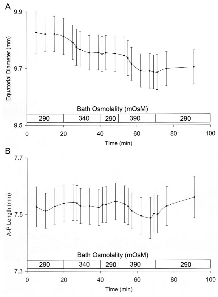
All experiments on rabbit lenses were performed over a tonicity range of −90 mOsM to +100 mOsM relative to the control osmolality of 290 mOsM. Increasing the bath tonicity with 50 mM sucrose produced a discernable reduction in ED by 0.6% from 9.81 to 9.76 mm without a detectable effect on the A-P length (Table 1). This 0.05 mm ED reduction represents an estimate of the maximal change that occurred given that changes ≥ 0.1 mm are assuredly measured. The control bathing solution was then reintroduced, which had no effect on the ED or A-P length (Table 1), and the lenses were then exposed to a second hypertonic challenge by the replacement of the medium with Tyrode’s solution containing 100 mM sucrose. This larger tonicity shift reduced ED by 1.2% relative to its control value and also reduced the A-P length by 0.7% relative to its respective control, from 7.55 to 7.50 mm (Table 1). These changes occurred within 10–20 min of the increase in bath osmolality. Fig. 2 depicts the time course of the measured changes for the 8 lenses listed in Table 1. The subsequent reintroduction of the control bath (now representing a 100 mOsM hypotonic shift from 390 to 290 mOsM) did not elicit measurable recoveries in the ED or A-P length (Table 1). Thus, the induced osmotically driven changes in volume were irreversible in the isolated rabbit lens within the time frame of our observations. Upon the return to the initial control conditions, the lens had acquired a different shape.
Fig. 2.
Time course of the changes in ED (panel A) and A-P length (panel B) upon initially introducing hypertonic media. A plot of the data from the lenses in Table 1. The tonicity shifts were accomplished by a total replacement of the lens bathing solutions with fresh media having a modified osmolality as indicated. During this replacement, which typically took about 3–5 min, the running time clock for the experiment was suspended and restarted as soon as the lens was covered with the fresh replacement solution. As such, the x-axis indicates the time of incubation in each osmolality.
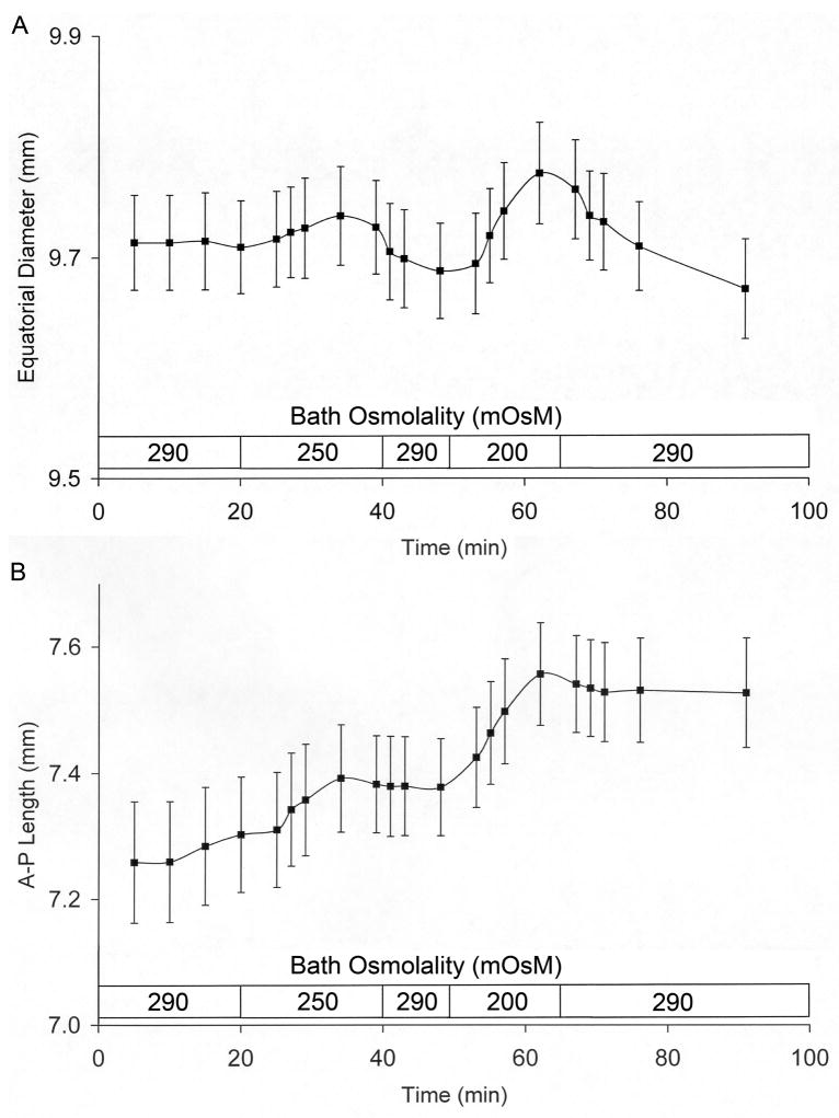
The imposition of hypotonic conditions as the initial experimental perturbation had proportionally larger effects on the A-P length than on ED (Table 2). The lenses were handled as above, except that the tonicity of the bathing solution was sequentially reduced (−40 mOsM), restored (+40 mOsM), reduced a second time (−90 mOsM) and restored again (+90 mOsM). The initial −40 mOsM shift increased the A-P length by ≈ 1.1% without markedly affecting ED (Table 2). In the presence of the larger tonicity shift (−90 mOsM), ED increased by ≈ 1.1% while the A-P length increased by ≈ 3.2%. ED reverted to its control value upon the reintroduction of the control medium at the end of the experiments, whereas the A-P length was unaffected by this hypertonic shift (i.e., from 200 to 290 mOsM). The time course of these measured changes from the 8 lenses in Table 2 are plotted in Fig. 3.
Table 2.
Effect of Hypotonic Conditions on the Equatorial Diameter (ED) and Length of the Anterior-Posterior (A-P) Axis of Isolated Rabbit Lenses.
| Equatorial Diameter (mm) | A-P Length (mm) | |||||||||
|---|---|---|---|---|---|---|---|---|---|---|
| Lens No. | Control (290 mOsM) | Tonicity Reduced (250 mOsM) | Restore Control Solution (290 mOsM) | Tonicity Reduced (200 mOsM) | Restore Control Solution (290 mOsM) | Control (290 mOsM) | Tonicity Reduced (250 mOsM) | Restore Control Solution (290 mOsM) | Tonicity Reduced (200 mOsM) | Restore Control Solution (290 mOsM) |
| Exp 17 | 9.80 | 9.80 | 9.76 | 9.84 | 9.72 | 7.54 | 7.60 | 7.56 | 7.69 | 7.66 |
| Exp 20 | 9.93 | 9.98 | 9.93 | 10.03 | 9.93 | 7.33 | 7.42 | 7.40 | 7.63 | 7.60 |
| Exp 21 | 9.70 | 9.75 | 9.68 | 9.78 | 9.67 | 7.09 | 7.22 | 7.20 | 7.43 | 7.41 |
| Exp 22 | 9.66 | 9.74 | 9.63 | 9.77 | 9.63 | 6.96 | 7.08 | 7.15 | 7.34 | 7.38 |
| Exp 23 | 9.69 | 9.72 | 9.69 | 9.77 | 9.68 | 7.50 | 7.62 | 7.53 | 7.70 | 7.58 |
| Exp 24 | 9.55 | 9.55 | 9.52 | 9.60 | 9.50 | 7.70 | 7.75 | 7.75 | 7.97 | 8.00 |
| Exp 29 | 9.74 | 9.75 | 9.72 | 9.78 | 9.67 | 7.17 | 7.26 | 7.26 | 7.39 | 7.33 |
| Exp 30 | 9.60 | 9.62 | 9.58 | 9.64 | 9.56 | 7.12 | 7.19 | 7.18 | 7.31 | 7.29 |
| Mean: | 9.71 | 9.74 | 9.69 | 9.78* | 9.67** | 7.30 | 7.39*** | 7.38*** | 7.56Δ | 7.53Δ |
| (n): | 8 | 8 | 8 | 8 | 8 | 8 | 8 | 8 | 8 | 8 |
| SEM: | 0.04 | 0.05 | 0.04 | 0.05 | 0.05 | 0.09 | 0.09 | 0.08 | 0.08 | 0.08 |
Lenses were handled in a manner similar to that described in the Table 1 legend, except that the tonicity of the bathing solution was sequentially reduced, restored, reduced a second time (to 200 mOsM), and restored again. Compiled values carry an insignificant figure. Changes ≥ 0.1 mm could be readily detected by the method used.
ED value significantly greater by ≈ 0.9% (P< 0.02, as two-tailed paired data), or
less by ≈ 1.1 % than its immediate antecedent (P< 0.001, as two-tailed paired data).
Value significantly greater by ≈ 1.1 % than the initial baseline value, 7.30 (P< 0.001, as two-tailed paired data).
Value significantly greater by ≈ 3.2 % than the initial baseline value, 7.30 (P< 0.001, as two-tailed paired data).
Fig. 3.
Time course of the changes in ED (panel A) and A-P length (panel B) upon initially introducing hypotonic media. A plot of the data from the lenses in Table 2. The x-axis indicates the time of incubation in each osmolality.
Overall, hypertonic shifts elicited larger effects on ED than the A-P length, whereas the converse occurs in hypotonic media. These experiments indicated that the lens does not change its shape in a uniform manner when challenged osmotically. Moreover, the ratio of the A-P length to the ED can be considered a measure of circularity, and this ratio increased more prominently in lenses exposed to hypotonic media than those under hypertonic conditions (Table 3). For example, with the more extreme −90 mOsM hypotonic solution the A-P length to ED ratio of 0.773 is 2.8% larger than the initial control ratio of 0.752 (Table 3). This increase is primarily due to the larger change in the A-P length relative to the change in ED.
Table 3.
Effects of Anisotonic Conditions on the A-P length to ED ratio of Isolated Rabbit Lenses
| Hypertonic Shifts | Hypotonic Shifts | ||||||||||
|---|---|---|---|---|---|---|---|---|---|---|---|
| Lens No. | Control (290 mOsM) | Sucrose 50 mM (340 mOsM) | Restore Control Solution (290 mOsM) | Sucrose 100 mM (390 mOsM) | Restore Control Solution (290 mOsM) | Lens No. | Control (290 mOsM) | Tonicity Reduced (250 mOsM) | Restore Control Solution (290 mOsM) | Tonicity Reduced (200 mOsM) | Restore Control Solution (290 mOsM) |
| Exp 13 | 0.747 | 0.759 | 0.757 | 0.758 | 0.761 | Exp 17 | 0.770 | 0.776 | 0.775 | 0.781 | 0.788 |
| Exp 14 | 0.774 | 0.777 | 0.778 | 0.778 | 0.778 | Exp 20 | 0.738 | 0.743 | 0.745 | 0.760 | 0.765 |
| Exp 15 | 0.745 | 0.751 | 0.750 | 0.751 | 0.754 | Exp 21 | 0.731 | 0.741 | 0.744 | 0.760 | 0.766 |
| Exp 16 | 0.757 | 0.764 | 0.766 | 0.770 | 0.766 | Exp 22 | 0.721 | 0.727 | 0.742 | 0.751 | 0.766 |
| Exp 25 | 0.788 | 0.790 | 0.792 | 0.792 | 0.797 | Exp 23 | 0.774 | 0.784 | 0.777 | 0.789 | 0.783 |
| Exp 26 | 0.812 | 0.809 | 0.816 | 0.811 | 0.821 | Exp 24 | 0.807 | 0.812 | 0.814 | 0.830 | 0.842 |
| Exp 27 | 0.759 | 0.759 | 0.761 | 0.757 | 0.763 | Exp 29 | 0.736 | 0.745 | 0.747 | 0.755 | 0.758 |
| Exp 28 | 0.766 | 0.768 | 0.772 | 0.771 | 0.772 | Exp 30 | 0.742 | 0.747 | 0.749 | 0.758 | 0.762 |
| Mean: | 0.769 | 0.772* | 0.774* | 0.774* | 0.777* | Average: | 0.752 | 0.759** | 0.762** | 0.773** | 0.779** |
| (n): | 8 | 8 | 8 | 8 | 8 | (n): | 8 | 8 | 8 | 8 | 8 |
| SEM: | 0.008 | 0.007 | 0.008 | 0.007 | 0.008 | SEM: | 0.010 | 0.010 | 0.009 | 0.009 | 0.010 |
The ratio of the AP-length to the ED of the lenses presented in Tables 1 and 2 are compiled as a measure of the changes in lens “circularity” under the imposed anisotonic conditions.
Value significantly greater than the initial control value at beginning of experiment, 0.769 (P< 0.03, as paired data).
Value significantly greater than its respective antecedent (P< 0.001, as paired data).
In contrast, in the more extreme +100 mOsM hypertonic solution, lenses exhibited a “circularity” ratio of 0.774, a value only 0.65% larger than the respective initial control value of 0.769; indicating that the decrease in ED under hypertonic conditions was not sufficient to dramatically increase the “circularity” of the lens (Table 3). Furthermore, the A-P length to ED ratio was relatively constant in the set of experiments with the hypertonic shifts as the initial test perturbation (Table 3, left side), while the set of experiments in which the lenses were exposed initially to a hypotonic solution (Table 3, right side) resulted in the “circularity” increasing gradually during the course of the experiments.
Osmotically Evoked Changes in Rabbit Lens Volume
Table 4 compiles the calculated volumes of the rabbit lenses detailed above in Tables 1 and 2 throughout the sequential changes in bath tonicity. The volumes of lenses exposed to a hypertonic shift as the initial experimental perturbation are presented on the left side of Table 4, and those lenses initially tested with hypotonic media are on the right. Overall, the general protocol of 4 sequential changes in bath tonicities resulted in lower lens volumes in those lenses initially exposed to a hypertonic perturbation and larger volumes in those lenses initially exposed to hypotonic shifts. A recovery from the experimentally induced volume changes to the initial control volumes was not seen within the time course of the present experiments.
Table 4.
Effects of Anisotonic Conditions on the Calculated Volume (mm3) of Isolated Rabbit Lenses
| Hypertonic Shifts | Hypotonic Shifts | ||||||||||
|---|---|---|---|---|---|---|---|---|---|---|---|
| Lens No. | Control (290 mOsM) | Sucrose 50 mM (340 mOsM) | Restore Control Solution (290 mOsM) | Sucrose 100 mM (390 mOsM) | Restore Control Solution (290 mOsM) | Lens No. | Control (290 mOsM) | Tonicity Reduced (250 mOsM) | Restore Control Solution (290 mOsM) | Tonicity Reduced (200 mOsM) | Restore Control Solution (290 mOsM) |
| Exp 13 | 402 | 396 | 396 | 390 | 391 | Exp 17 | 366 | 364 | 359 | 373 | 368 |
| Exp 14 | 393 | 390 | 388 | 379 | 380 | Exp 20 | 362 | 380 | 379 | 393 | 389 |
| Exp 15 | 354 | 350 | 348 | 343 | 341 | Exp 21 | 347 | 353 | 350 | 366 | 362 |
| Exp 16 | 353 | 349 | 352 | 344 | 345 | Exp 22 | 351 | 359 | 353 | 367 | 361 |
| Exp 25 | 372 | 366 | 365 | 358 | 360 | Exp 23 | 355 | 365 | 355 | 368 | 359 |
| Exp 26 | 373 | 370 | 370 | 362 | 363 | Exp 24 | 352 | 351 | 347 | 362 | 356 |
| Exp 27 | 354 | 350 | 350 | 344 | 343 | Exp 29 | 350 | 359 | 352 | 361 | 361 |
| Exp 28 | 375 | 370 | 368 | 363 | 364 | Exp 30 | 337 | 337 | 334 | 350 | 344 |
| Mean: | 372 | 368* | 367 | 360* | 361 | Average: | 352 | 358** | 354* | 367** | 362* |
| (n): | 8 | 8 | 8 | 8 | 8 | (n): | 8 | 8 | 8 | 8 | 8 |
| SEM: | 6.5 | 6.4 | 6.2 | 6.1 | 6.3 | SEM: | 3.2 | 4.4 | 4.5 | 4.4 | 4.5 |
Volumes of the lenses shown in Tables 1 and 2 were calculated for each incubation condition using the presented ED and A-P length plus the CSA and center of mass that were measured from the lens profiles as briefly described in Methods and in detail in Gerometta et al., (2007). Volume changes greater than 1% could be readily detected by the method used. Hypertonic solutions of +50 mOsM and +100 mOsM decreased rabbit lens volumes by 1.1% and 3.2% relative to control baseline value, 372. Hypotonic solutions of −40 mOsM and −100 mOsM increased rabbit lens volumes by 1.7% and 4.3% relative to the baseline value, 352; lens volume declined by 1.4% upon restoring the control solution at the end of the experiment.
Value significantly less than its respective antecedent (P< 0.001, as paired data).
Value significantly greater than its respective antecedent (P< 0.025, as paired data).
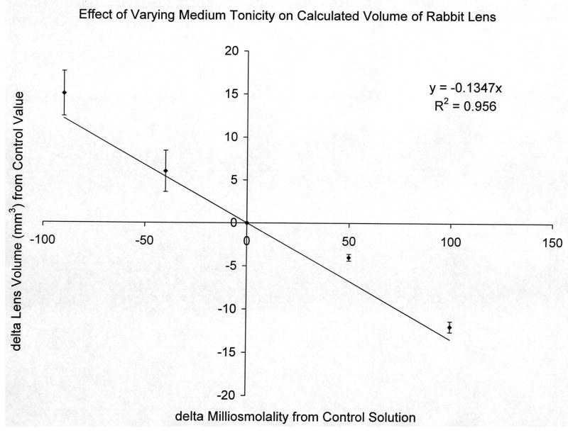
Although the induced volume changes were small, a plot of the data in Table 4 showing the changes in lens volume as a function of the changes in bath tonicity illustrated that rabbit lens volume responded in a linear fashion to the imposed osmotic challenges (Fig. 4), indicating that within a 10–20 min time frame, the whole lens responded as an osmometer over a −90 to +100 mOsM tonicity range.
Fig. 4.
A plot of the changes in rabbit lens volume as a function of the changes in bath osmolality from the values in Table 4.
Osmotically Evoked Changes in Cow Lens Shape and Volume
The physical parameters of cow lenses were measured and analyzed as done with rabbit lenses, but the incubation protocols were different. Two general approaches were done – 1) monitor the control parameters for 45 min followed by an additional 45 min of monitoring after a tonicity shift of either +150 mOsM (Table 5), or −100 mOsM (Table 6); and 2) placing the cow lens directly (at t= 0) in the observation chamber in a hypotonic medium of 190 mOsM (Fig. 5). The latter condition was done to illustrate the very gradual changes in the A-P length and ED dimensions, as well as in volume, exhibited by the cow lens when swelling. However, this latter protocol is not presented for the hypertonic condition because the cow lens was surprisingly refractory to increases in medium osmolality and very meager changes were obtained.
Table 5.
Effects of Hypertonic Conditions on Physical Parameters of Isolated Cow Lenses
| Control Solution (290 mOsM) | Hypertonic Solution (440 mOsM) | |||||||
|---|---|---|---|---|---|---|---|---|
| Lens No. | A-P Length (cm) | ED (cm) | A-P/ED | Lens Volume (cm3) | A-P Length (cm) | ED (cm) | A-P/ED | Lens Volume (cm3) |
| Exp 10 | 1.21 | 1.71 | 0.708 | 1.72 | 1.21 | 1.67 | 0.725 | 1.72 |
| Exp 13 | 1.22 | 1.83 | 0.667 | 2.08 | 1.22 | 1.80 | 0.678 | 2.05 |
| Exp 14 | 1.17 | 1.69 | 0.692 | 1.74 | 1.17 | 1.68 | 0.696 | 1.72 |
| Exp 15 | 1.30 | 1.81 | 0.718 | 2.17 | 1.30 | 1.79 | 0.726 | 2.16 |
| Exp 16 | 1.22 | 1.71 | 0.713 | 1.85 | 1.22 | 1.69 | 0.722 | 1.83 |
| Mean: | 1.22 | 1.75 | 0.700 | 1.91 | 1.22 * | 1.73 | 0.710** | 1.90* |
| (n): | 5 | 5 | 5 | 5 | 5 | 5 | 5 | 5 |
| SEM: | 0.021 | 0.029 | 0.009 | 0.091 | 0.021 | 0.028 | 0.010 | 0.089 |
Cow lenses were incubated for 45 minutes in control physiological solution (290 mOsM). The tabulated, control parameters were measured as described in Methods from photographs taken immediately prior to replacing the control medium with a +150 mOsM hypertonic solution. Photographs for the experimental condition were taken 45 minutes after the solution change (t= 90 min of the experiment). Changes greater than 0.01 cm in the A-P length and ED dimensions, and volume changes greater than 1% could be readily detected by the method used. Cow lens ED deceased by 1.1% and lens volume by 0.5% in the hypertonic medium. The A-P to ED ratio increased by 1.4%.
Value significantly less than its respective control value (P< 0.05, as paired data).
Value significantly greater than its respective control value (P< 0.01, as paired data).
Table 6.
Effects of Hypotonic Conditions on Physical Parameters of Isolated Cow lenses
| Control Solution (290 mOsM) | Hypotonic Solution (190 mOsM) | |||||||
|---|---|---|---|---|---|---|---|---|
| Lens No. | A-P Length (cm) | ED (cm) | A-P/ED | Lens Volume (cm3) | A-P Length (cm) | ED (cm) | A-P/ED | Lens Volume (cm3) |
| Exp 1 | 1.29 | 1.84 | 0.701 | 2.20 | 1.33 | 1.84 | 0.723 | 2.28 |
| Exp 2 | 1.22 | 1.81 | 0.674 | 2.01 | 1.24 | 1.81 | 0.685 | 2.08 |
| Exp 5 | 1.25 | 1.72 | 0.727 | 1.89 | 1.28 | 1.72 | 0.744 | 1.95 |
| Exp 6 | 1.34 | 1.89 | 0.709 | 2.46 | 1.38 | 1.89 | 0.730 | 2.50 |
| Exp 7 | 1.27 | 1.73 | 0.734 | 1.95 | 1.31 | 1.75 | 0.749 | 2.04 |
| Mean: | 1.27 | 1.80 | 0.709 | 2.10 | 1.31* | 1.80 | 0.726* | 2.17* |
| (n): | 5 | 5 | 5 | 5 | 5 | 5 | 5 | 5 |
| SEM: | 0.020 | 0.032 | 0.011 | 0.10 | 0.024 | 0.031 | 0.011 | 0.10 |
Cow lenses were incubated for 45 minutes in control physiological solution (290 mOsM). The tabulated, control parameters were measured as described in Methods from photographs taken immediately prior to replacing the control medium with a −100 mOsM hypotonic solution. Photographs for the experimental condition were taken 45 minutes after the solution change (t= 90 min of the experiment). Changes greater than 0.01 cm in the A-P length and ED dimensions, and volume changes greater than 1% could be readily detected by the method used. Cow lens A-P length increased by 3.1%, the A-P to ED ratio by 3.8%, and the lens volume by 3.3% in the hypotonic medium.
Value significantly greater than its respective control value (P< 0.001, as paired data).
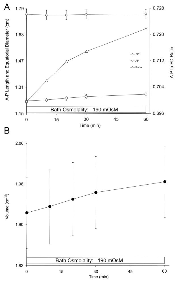
Fig. 5.
A) Plot of the measured A-P length (open circles) and equatorial diameter (open diamonds) of cow lenses bathed in a hypotonic solution over the course of an hour. The calculated A-P length-to-ED ratio (open triangles) increased solely due to an increase in the A-P length. Measured values are the means ± SE’s from 5 lenses.
B) Plot of the calculated lens volume of the 5 lenses in Plot A, using the same lens profiles that were digitally captured for the A-P length and ED measurements plus the measured CSA and center of mass that were also obtained from the images. See details in Gerometta et al. (2007).
Hypertonic solutions of +100 mOsM did not affect on the measured parameters (data not shown), although a mild cloudiness of the superficial lens surface could be clearly seen in the photographs taken at the point of the solution change. This indicates that the lens was affected by the tonicity perturbation, but neither its shape nor volume changed. (No opacification of lenses was ever observed under hypotonic conditions given the short duration of the experiments and the limited volume increases recorded.) Exposing the cow lenses to hypertonic solutions of +150 mOsM elicited a more conspicuous cloudiness at the lens surface, a reduction in ED of 1.1% (to 1.73 mm; Table 5) and a volume decline of 0.5% (to 1.90 cm3; Table 5). No measurable decreases in A-P length were detected.
In contrast, hypotonic conditions evoked conspicuous changes in the dimensional parameters of the cow lens. As shown in Table 6, a 3.3% increase in lens volume (to 2.17 cm3) was obtained following 45 min of exposure to the −100 mOsM hypotonic bath. This change concurred with a significant 3.1% increase in the A-P length, and no detectable change in ED (Table 5). As shown in Tables 5 and 6, the A-P to ED ratio increased under both the hypertonic and hypotonic conditions, showing that the lens “circularity” is augmented osmotically irrespective of the direction of the tonicity shift. However, the increase in circularity was proportionally larger when the lenses swelled (Table 6).
The cow lens experiments that entailed isolating the lenses with control solution (290 mOsM) and then placing the lenses in the observation chamber from t= 0 with an hypotonic medium (190 mOsM) is essentially a permutation of the conditions presented in Table 6. In this set of experiments, the lenses were photographed over the course of 1 hour and the A-P length, ED, A-P to ED ratio, and calculated volume were plotted (Fig. 5). Although hypotonic shifts produced more prominent changes in the lens dimensional parameters than did hypertonic conditions, the cow lens dimensions changed very gradually as illustrated in the figure. In this set of cow lenses, a 3% volume increase from 1.92 ± 0.07 cm3 at t= 0 to 1.98 ± cm3 at t= 60 min (n= 5, P< 0.001 as paired data) occurred simultaneously with a 4% increase in the A-P length (from 1.22 ± 0.013 cm to 1.27 ± 0.012 cm, n=5, P< 0.001), results similar to those obtained with the lenses in Table 6. As before, the ED did not change (P> 0.9, as paired data).
Summarizing our overall results with the lenses of both species, anisotonic conditions produced an increase in circularity irrespective of the direction of the tonicity change. Larger increases in circularity occurred with the hypotonic conditions, under which the increase in A-P length was the predominant shape change. This parameter increased 0.26 mm (a 3.5% change) in rabbit lenses, which simultaneously gained 15 μl (a 4.3% increase), in −90 mOsM solution within 20 min of exposure to the lower tonicity, while in cow lenses the increases were 0.40 mm (a 3.1% change) and 70 μl (a 3.3% increase) in −100 mOsM solution within 45 min of exposure. These changes were proportionally similar between the two species of lenses.
Hypertonic solutions did not induce a decrease in the A-P length as occurs during dis-accommodation when the lens gets flatter. Instead the ED was primarily reduced. Rabbit lenses lost 12 μl (a 3.2% loss) in tandem with a 0.11 mm ED reduction (a 1% decrease) in +100 mOsM solution within 20 min, while cow lenses lost 10 μl (a 0.5% loss) along with a 0.20 mm ED reduction (a 1% decrease) in +150 mOsM solution within 45 min. In this comparison, a higher tonicity shift and time of exposure were required with the cow lens in order to obtain similar proportional changes in ED, and similar absolute changes in volume, but the larger mass of the cow lens resulted in a markedly smaller, calculated percentage change in the fluid lost under the hypertonic conditions.
Discussion
The lens is a quasi-spherical syncytium comprised entirely of epithelial cells at various stages of differentiation with localized transport properties (Mathias, Kistler and Donaldson, 2007). Metabolism occurs throughout the lens enabled by a hypothetical internal circulation system driven by ionic pumps, channels and transporters (Mathias et al., 2007; Donaldson et al., 2008). Many of the transport elements that are proposed to underpin the internal lens circulation system are recognized as having pivotal roles in volume regulation in single cells (e.g., red blood cells, cultured lens epithelial cells; Diecke and Beyer-Mears, 1997; Reeves et al., 1998), and presumably contribute to the steady-state volume control of lenses under physiological conditions (Donaldson et al., 2008), as well as lens volume regulation in lenses perturbed by anisotonic media in vitro (Patterson and Fournier, 1976; Patterson, 1981).
With intact lenses, we have observed within minutes of an osmotic challenge the activation of the epithelial Na+-K+-2Cl− cotransport activity, and of lens epithelial K+ conductances that are volume responsive (Alvarez et al., 1991, 2001). This suggests that the lens epithelial cells, and perhaps the most superficial equatorial fibers (Donaldson et al., 2008), may transport sufficient solute to maintain their volumes in the face of extreme tonicity shifts. Simultaneously, the osmolality of the bulk of the lens water requires several hours to equilibrate with that of the altered external bath (Cotlier et al., 1968). It thus seems plausible that the intact lens cannot recover its original volume until the bulk of the lens has the same osmolality as that of the bathing medium. This may explain 1) the report that rat lenses required 1 to 2 days of incubation in organ culture (with Medium 199) to restore their original volume after undergoing osmotic swelling (Patterson and Fournier, 1976), and 2) the fact that we did not observe lens volume restoration in the course of this study in either rabbit or cow lenses.
Although we obtained linear changes in rabbit lens volumes within 20 min of the imposed osmotic challenges (Fig. 4), a finding similar to that of Kinoshita et al. (1965) and Cotlier et al. (1968), clearly the lens is not a “perfect osmometer” given the presence of volume regulatory mechanisms, which probably contributed to the relatively small volume changes that we observed in the presence of large tonicity shifts and the limited time of exposure that the rabbit lenses were bathed by anisotonic baths.
The majority of the rabbit lens experiments were conducted in media slightly hypertonic (+50, +100 mOsM) or hypotonic (−40, −90 mOsM) to the lens. Earlier, Kinoshita et al. (1965) observed that the rabbit lens responds as an osmometer through a limited range of tonicities. They demonstrated that changes in lens water content depended linearly upon the tonicity differences from the isotonic state when lenses were exposed to osmolalities differing from the control by −64 to +32 to mOsM. In that work the water contents were determined following 21 hours of anisotonic exposure, but some experiments were done at 4 hours. Our rabbit lens experiments measured changes in lens volume upon 10–20 min of anisotonic exposure, and found linear changes in volume as a function of the changes in bath tonicity over a somewhat larger range. We had initially chosen these larger tonicity shifts to be certain of the magnitude of the changes in the measured A-P length and ED.
Cotlier et al. (1968) also advanced the concept of the lens as an osmometer and plotted changes in water content of rabbit lenses incubated in anisotonic media on an hourly basis for a period of 4 hours. Lenses incubated in hypotonic media of −32, −64 and −260 mOsM gradually gained 5%, 14% and ≈65% of their original water content, while lenses bathed in hypertonic media of +33 and +66 mOsM lost 5% and 11% of their water content, respectively. Moreover, these authors also determined the intralenticular osmolalities of rabbit lenses incubated in the above anisotonic solutions. Their results indicated that the water contents of the lenses were gradually altered until the lenses were in equilibrium with the media, which could occur within 4 to 24 hours depending upon the extent of the tonicity shift.
The small changes in rabbit lens volume that we calculated for lenses exposed to tonicity shifts of −90 to +100 mOsM for 10–20 min (Fig. 4 and Table 4) could be expected to be larger upon prolonged incubation under these anisotonic conditions. Presumably, extended incubations with these tonicity shifts may not affect the linearity of the plot (Fig. 4), but rather steepen its slope. Any putative volume regulation by the whole lens might not be expected until the lens osmolality is fully equilibrated with the media. In short, the magnitude of the tonicity shift, the extent of time the lens is exposed to the altered osmolality, and the relative size of the lens may all affect the dynamics of volume regulation by the whole lens.
In this work, we primarily focused on the initial changes in lens shape and volume in response to the tonicity shifts and examined the effects of restoring the control conditions in some experiments. The latter maneuver did not result in a restoration of the original lens shape or volume, in contrast to the situation in accommodation under which lens shape, and possibly volume, are fully reversible.
Secondly, we aimed to determine whether the volume changes were symmetrical, i.e., uniform in a way that the shape of the lens was preserved, or asymmetrical as indicated by Zhang and Jacob (1996) for the bovine lens under hypotonic conditions, under which the lenses only increased the A-P length. Zhang and Jacob (1996) also reported a restoration of the A-P length towards the control value while the lens was maintained in the hypotonic medium, a result that we could not substantiate. However, the shape changes under hypotonic and hypertonic conditions are distinct.
Our experiments used a method that we developed (Gerometta et al., 2007; Zamudio et al., 2008) that allows for the determination of rapid changes in lens shape and volume. From this approach, the following observations were evinced.
With rabbit lenses, the predominant effect of hypertonic solution is on the ED, with markedly smaller effects on the A-P length, while the latter parameter is primarily affected under hypotonic conditions. Both conditions tend to increase lens “circularity”.
With cow lenses, hypertonic conditions solely affected ED, while hypotonic conditions solely affected the A-P length. The absence of detectable changes with cow lenses in one of the 2 parameters under our conditions was not due to a lack of resolution in the methods used. Changes in the responsive parameter, and in the calculated lens volumes, could be obtained with cow lenses within minutes of a tonicity change. But overall, cow lens shapes and volumes were less responsive than rabbit lenses to changes in bath tonicities; hence, experiments with the bovine specimen involved larger changes in bath osmolality.
It seems likely that the interdigitations of the fibers at the lens sutures, and the fact that the fibers are under tension by a restrictive capsule, probably influences the A-P length more than the ED. The former parameter increases and decreases (freely) during accommodation and dis-accommodation respectively, and an analogous increase in A-P length was obtained under hypotonic conditions.
During the accommodation phase, which is a totally passive phenomenon, the lens becomes rounder (i.e., there is an increase in circularity), and its volume may increase (Gerometta et al., 2007). Similarly, increases in circularity occur when lenses swell under hypotonic conditions primarily because the distance between the poles along the A-P axis lengthens more than the ED. However, in rabbit lens experiments, restoring the control osmolality does not revert the A-P length to its control state, while the ED, which changed less extensively, is restored. These observations suggest that the hypotonic swelling may have irreversibly altered the physical structures of the fibers at the sutures, at least within the time frame of the current experiments.
Alternatively, the Na+-K+ pumps and K+-Cl− cotransporters necessary for volume regulatory decrease (RVD) appear to have a higher activity near the lens equator in both the epithelium and superficial fibers (Mathias et al., 2007; Donaldson et al., 2008). An active RVD process concentrated at the rabbit lens equator may have attenuated the increase in ED under hypotonic conditions, and contributed to the restoration of ED upon returning to the control medium while the lens volume only declined by a negligible 1.4% (Table 4). Likewise, the absence of an effect by hypotonicity on the cow lens ED could also be due to a very active equatorial RVD response, or perhaps the different internal suture architecture than in the rabbit lens.
As characterized in detail by Kuszak et al. (2006), the suture architecture is critical for the spontaneous change in lens shape that occurs in response to the altered tension exerted by the ciliary muscle during the accommodation process. It appears that the terminal regions of the fiber cells have a “spring-like” quality that would tend to increase the A-P length, a tendency counterbalanced by the tension of the capsule. As this tension is relaxed during accommodation, the A-P length increases. Moreover, freeing the lens from its zonular attachments enables the lens to exhibit its maximal spontaneous increase in A-P length, suggesting that the “spring-like forces” within the fibers had completely dissipated. Hypotonically evoked swelling can then apparently produce a further extension of the fiber cell structure at the sutures to increase the A-P length more than the ED. This indicates that lens swelling is not symmetrical, but rather, a conformational change apparently consistent with the architecture of the lens that enables spontaneous changes in lens shape during accommodation. Conversely, hypertonically induced shrinkage occurs primarily due to a reduction in ED with proportionally smaller changes in A-P length in the rabbit lens, and no change in A-P length in the lens of the cow.
Rabbit lenses exposed to hypertonic conditions exhibit an activation of the epithelial Na+-K+-2Cl− cotransport activity, which is found throughout the entire lens epithelium from the anterior pole to the equator (Alvarez et al., 2001). This Na+-linked solute carrier is involved in regulatory volume increase (RVI) mechanisms in several cell systems (Russell, 2000), and may have such a role in the lens. If so, it is unclear as to how such activity might have maintained the A-P length, which was less responsive than ED to the hypertonic medium, given the uniform functional expression of the cotransporter in the epithelium, and the relatively high expression of NKCC1 in the equatorial cortex (Alvarez et al., 2001). While NKCC1 may have contributed towards the preservation of lens volume during our experiments, another explanation appears necessary as to why lens “circularity” increased under hypertonic conditions (i.e., ED decreased but A-P length was not markedly affected during osmotically induced lens shrinkage).
In short, we have demonstrated a method to measure rapid changes in lens volume and showed that anisotonically elicited changes in lens shape are asymmetrical phenomena because the lens does not swell or shrink evenly in all directions upon exposure to an altered osmolality. Presumably, the lenticular architecture that buttresses the shape changes observed during accommodation, when the lens becomes rounder, also underpins some of the changes in lens shape that are evoked by hypotonicity. Volume regulatory mechanism may also come into play. However, the tonicity shifts used in the present study were too large to obtain volume and shape recoveries within the time frame of the experiments. This underlines the small range under which lens shape can be reversibly changed. Hypothetically, it appears possible that reversible shape changes may be obtained with shorter exposure to less extreme osmotic challenges. On the other hand, it could also be that osmotic forces deform the lens differently than the changes produced by the mechanical forces of accommodation. It seems possible that osmotic forces predestine the lens from recovering its initial shape in a reversible manner. Osmotic and mechanical forces may act on the lens in different ways.
Acknowledgments
This work was supported by National Eye Institute Grants EY00160 and EY01867, and by an unrestricted grant from Research to Prevent Blindness, Inc., New York, NY.
Footnotes
Publisher's Disclaimer: This is a PDF file of an unedited manuscript that has been accepted for publication. As a service to our customers we are providing this early version of the manuscript. The manuscript will undergo copyediting, typesetting, and review of the resulting proof before it is published in its final citable form. Please note that during the production process errors may be discovered which could affect the content, and all legal disclaimers that apply to the journal pertain.
Contributor Information
Chi-Wing Kong, Email: marcokongcw@gmail.com.
Rosana Gerometta, Email: rgerometta@yahoo.com.ar.
Lawrence J. Alvarez, Email: larry.alvarez@mssm.edu.
Oscar A. Candia, Email: oscar.candia@mssm.edu.
References
- Alvarez LJ, Candia OA, Turner HC, Polikoff LA. Localization of a Na+-K+-2Cl− cotransporter in the rabbit lens. Exp Eye Res. 2001;73:669–680. doi: 10.1006/exer.2001.1075. [DOI] [PubMed] [Google Scholar]
- Alvarez LJ, Wolosin JM, Candia OA. Contribution from a pH- and tonicity-sensitive K+ conductance to toad translens short-circuit current. Exp Eye Res. 1991;52:283–292. doi: 10.1016/0014-4835(91)90092-s. [DOI] [PubMed] [Google Scholar]
- Candia OA. Reply to “Letter to the editor: ‘Ocular lens does not change volume during accommodation’”. Am J Physiol Cell Physiol. 2007;293:C1729–C1730. doi: 10.1152/ajpcell.00266.2007. [DOI] [PubMed] [Google Scholar]
- Cotlier E, Kwan B, Beaty C. The lens as an osmometer and the effects of medium osmolarity on water transport, 86Rb efflux and 86Rb transport by the lens. Biochim Biophys Acta. 1968;150:705–722. doi: 10.1016/0005-2736(68)90060-6. [DOI] [PubMed] [Google Scholar]
- Diecke FP, Beyer-Mears A. A mechanism for regulatory volume decrease in cultured lens epithelial cells. Curr Eye Res. 1997;16:279–288. doi: 10.1076/ceyr.16.4.279.10693. [DOI] [PubMed] [Google Scholar]
- Donaldson PJ, Chee KS, Lim JC, Webb KF. Regulation of lens volume: implications for lens transparency. Exp Eye Res. 2009;88:144–150. doi: 10.1016/j.exer.2008.05.011. [DOI] [PubMed] [Google Scholar]
- Gerometta R, Zamudio AC, Escobar DP, Candia OA. Volume change of the ocular lens during accommodation. Am J Physiol Cell Physiol. 2007;293:C797–804. doi: 10.1152/ajpcell.00094.2007. [DOI] [PubMed] [Google Scholar]
- Kinoshita JH, Merola LO, Hayman S. Osmotic Effects on the Amino Acid-Concentrating Mechanism in the Rabbit Lens. J Biol Chem. 1965;240:310–315. [PubMed] [Google Scholar]
- Kuszak JR. The ultrastructure of epithelial and fiber cells in the crystalline lens. Int Rev Cytol. 1995;163:305–350. doi: 10.1016/s0074-7696(08)62213-5. [DOI] [PubMed] [Google Scholar]
- Kuszak JR, Mazurkiewicz M, Jison L, Madurski A, Ngando A, Zoltoski RK. Quantitative analysis of animal model lens anatomy: accommodative range is related to fiber structure and organization. Vet Ophthalmol. 2006;9:266–280. doi: 10.1111/j.1463-5224.2006.00506.x. [DOI] [PubMed] [Google Scholar]
- Mathias RT, Kistler J, Donaldson P. The lens circulation. J Membr Biol. 2007;216:1–16. doi: 10.1007/s00232-007-9019-y. [DOI] [PubMed] [Google Scholar]
- Patterson JW. Lens volume regulation in hypertonic medium. Exp Eye Res. 1981;32:151–162. doi: 10.1016/0014-4835(81)90004-x. [DOI] [PubMed] [Google Scholar]
- Patterson JW, Fournier DJ. The effect of tonicity on lens volume. Invest Ophthalmol. 1976;15:866–869. [PubMed] [Google Scholar]
- Reeves RE, Sanchez-Torres J, Coca-Prados M, Cammarata PR. Expression of pI(Cln) mRNA in cultured bovine lens epithelial cells: response to changes in cell volume. Curr Eye Res. 1998;17:861–869. doi: 10.1076/ceyr.17.9.861.5140. [DOI] [PubMed] [Google Scholar]
- Russell JM. Sodium-potassium-chloride cotransport. Physiol Rev. 2000;80:211–276. doi: 10.1152/physrev.2000.80.1.211. [DOI] [PubMed] [Google Scholar]
- Zampighi GA, Eskandari S, Hall JE, Zampighi L, Kreman M. Micro-domains of AQP0 in lens equatorial fibers. Exp Eye Res. 2002;75:505–519. doi: 10.1006/exer.2002.2041. [DOI] [PubMed] [Google Scholar]
- Zhang JJ, Jacob TJ. Volume regulation in the bovine lens and cataract. The involvement of chloride channels. J Clin Invest. 1996;97:971–978. doi: 10.1172/JCI118521. [DOI] [PMC free article] [PubMed] [Google Scholar]


