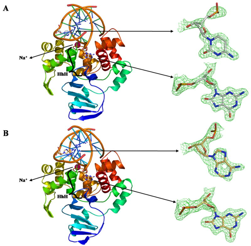
Figure 1. Overall fold of CacOggK222Q in complex with DNA containing A) 8-oxoG:C and B) 8-oxoG:A.

Ribbon diagrams of CacOggK222Q in complex with DNA containing A) 8-oxoG:C and B) 8-oxoG:A Proteins are colored according to the amino acid sequence going from cold blue to warm red from N- to C-terminal. A simulated annealing omit map (green) contoured at 3σ is shown for each of the estranged bases and 8-oxoG. The sodium atom is colored in pink in both panels and the HhH motif is labeled. All figures were prepared using PYMOL [38].
