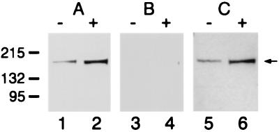
Figure 1.
Immunoblot analysis of AS3 protein induction in cellular extracts. Detection of AS3 in the LNCaP-FGC cell extract was carried out by using a rabbit polyclonal anti-AS3 antibody. The lysates used were from cells treated with 1 nM R1881 for 48 h (+) and cells treated for 48 h with vehicle only (−). The protein load was 35 μg/lane. (A) Uncompeted antibody; (B) peptide competition was performed with 50-fold excess of the specific C terminus peptide and (C) with a nonrelated N terminus peptide. The numbers (Left) indicate the molecular markers in kilodaltons; the arrow (Right) points to the AS3 band.

