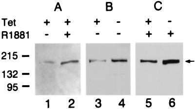
Figure 5.
Expression of AS3 protein in MCF7-AR1, and S9 cellular extracts. (A) Androgen induction of AS3 in MCF7-AR1 cells. Lane 1, cells treated with 100 pM estradiol for 48 h; lane 2, cells expressing the androgen-induced proliferative shutoff after 48 h exposure to 100 pM estradiol and 1 nM R1881. (B) Tet induction of AS3 in S9 cells. Lane 3, cells were grown in the presence of 100 pM estradiol and 1 μg/ml tet for 4 days; lane 4, cells were grown in the presence of 100 pM estradiol and with 1 μg/ml tet for the first 2 days and then without tet for an additional 2 days. (C) Tet induction of AS3 in S9 cells treated with R1881. Lanes 5 and 6, cells received the same treatment as in lanes 3 and 4, but they were also exposed to 10 nM R1881 for 4 days. The protein load was 35 μg/lane. The numbers (Left) indicate the molecular markers in kDa; the arrow (Right) points to the AS3 band.

