
Figure 1.
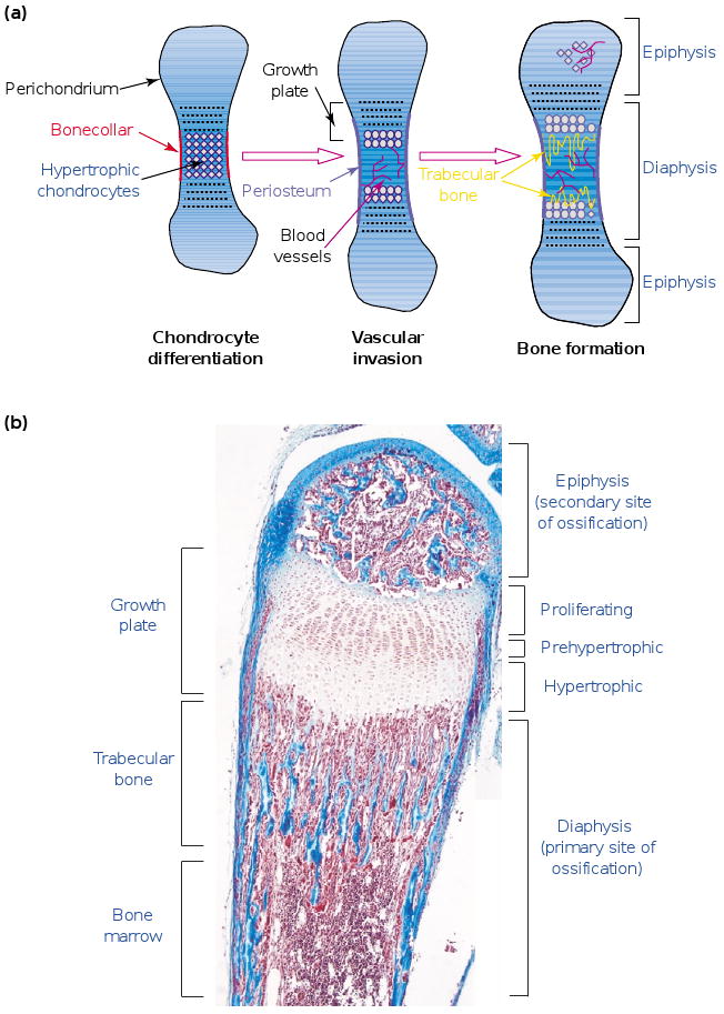
The process of endochondral ossification. (a) During endochondral ossification, mesenchymal cells differentiate into chondrocytes and lead to the formation of cartilage templates. Vascularization occurs around these templates, and osteoblasts differentiate around the central area in the bone collar. Chondrocytes in this central area differentiate into hypertrophic chondrocytes and allow vascular invasion. After complete differentiation, they die and extracellular matrix (ECM) remodeling is carried out by osteoclasts and chondroclasts recruited together with the blood vessels. This remodeling is necessary for trabecular-bone synthesis by osteoblasts precursors and for the formation of the bone-marrow cavity. (b) A section of a two-week-old mouse metatarsal stained with Masson trichrome. Chondrocytes proliferate and differentiate into prehypertrophic and hypertrophic chondrocytes. Endochondral ossification first takes place at the primary site at the center of the diaphysis, which allows formation of the two growth plates. The growth plates are ultimately responsible for the elongation of the long bones. Later, endochondral ossification occurs at a secondary site, in the epiphysis of the long bones. Scale bar represents 200 μm.