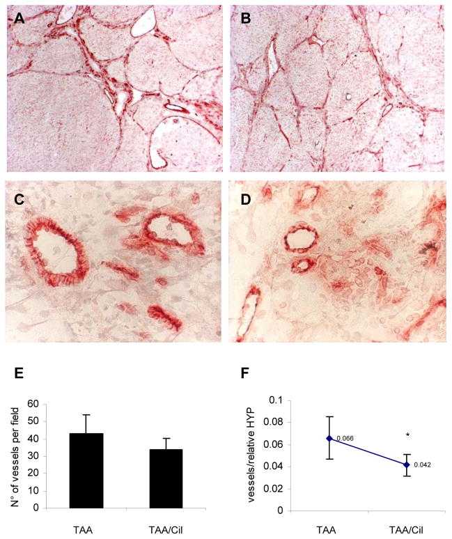
Fig.4. CD31 immunohistochemistry on livers of rats with TAA-induced fibrosis treated with/without Cilengitide.
(A) TAA-R (n=7, magnification 10x); (B) TAA-R + Cilengitide 30mg/kg/day 8w (n=8) (10x); (C) and (D), magnifications of (A) and (B), respectively (40x). (E) Quantification of angiogenesis. Calculation of CD31-positive vessels was performed in four areas from each liver section using light microscopy (magnification 10x) and is expressed as a number of vessels per field (means±SD). (F) Vessel number per fibrotic area is expressed as the ratio of vessels to relative hepatic HYP (means±SD), *p<0.05 vs TAA-R group.

