Figure 5.
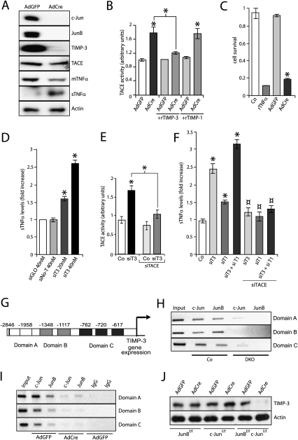
Increased TNFα shedding in keratinocytes conditionally deficient for JunB and c-Jun in vitro and upon TIMP-1 and TIMP-3 knockdown. (A) Western blot analysis of c-Jun, JunB, TIMP-3, TACE, and mTNFα in AdGFP- or AdCre-infected JunBf/f; c-Junf/f keratinocytes. For sTNFα, the sample was obtained from cell culture supernatant. Actin indicates equivalent loading. (B) Increased TACE activity in AdCre-infected keratinocytes (n = 3). The same extracts were assayed in the presence of recombinant TIMP-3 and TIMP-1. Data are shown as mean ± SD. (*) P < 0.05 (Student's t-test). (C) MTT assay performed with culture supernatant from AdGFP-infected (n = 3) or AdCre-infected (n = 3) keratinocytes. Keratinocyte medium and recombinant TNFα were used as negative (Co) and positive (rTNFα, 150 ng/mL) controls, respectively. Data are shown as mean ± SD. (*) P < 0.05 (Student's t-test). (D) sTNFα ELISA upon siRNA knockdown of mouse TIMP-3 (siT3) in wild-type keratinocytes. siGLO and siNo-T were used as transfection and target specificity controls, respectively. (E) Increased TACE activity in wild-type keratinocytes upon TIMP-3 knockdown (siT3). TACE siRNA (siTACE) inhibited the increase in TACE activity. Data are shown as mean ± SD. (*) P < 0.05 (Student's t-test). (F) Increased sTNFα shedding as measured by ELISA upon siRNA knockdown of mouse TIMP-1 (siT1), TIMP-3 (siT3), and TIMP-1/TIMP-3 (siT1 + siT3) in wild-type keratinocytes. The increased shedding was inhibited upon concomitant knockdown of TACE (siTACE). Data are shown as mean ± SD. (*) P < 0.05 (Student's t-test). (G) Schematic illustration of the mouse TIMP-3 promoter showing the six putative AP-1-binding sites. Due to ChIP constraints, the six sites were grouped into three different domains that could be amplified by different sets of primers. In the right panel, ChIP and PCR amplification demonstrates c-Jun and JunB binding to the TIMP-3 promoter in epidermal samples from Co, but not from DKO, pups at P1. When JunB and c-Jun were expressed, binding was detected in all three domains. (H) ChIP analysis of c-Jun and JunB binding to the TIMP-3 promoter in Co- and DKO-derived keratinocytes. (I) ChIP analysis of c-Jun and JunB binding to the TIMP-3 promoter in cultured AdGFP- and AdCre-infected JunBf/f; c-Junf/f keratinocytes. Isotype IgG was used as a control (IgG). (J) TIMP-3 Western blot analysis of AdGFP or AdCre JunBf/f, c-Junf/f or JunBf/f; c-Junf/f infected keratinocytes. Actin indicates equivalent loading.

