
Figure 1.
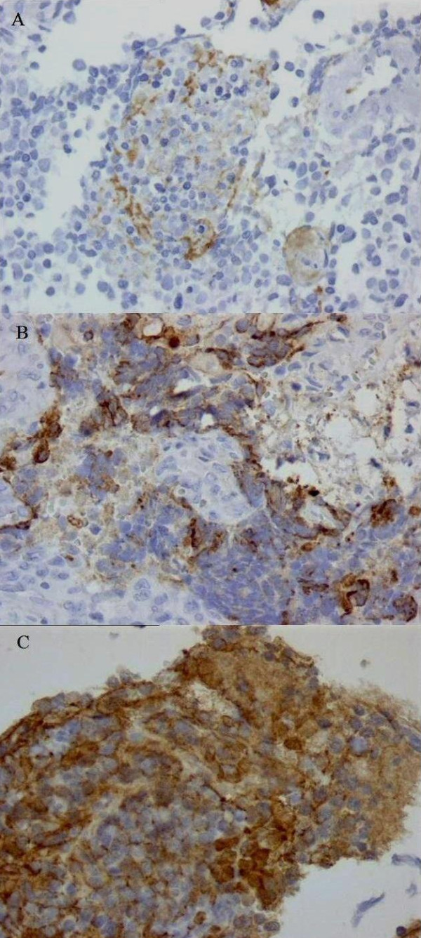
Immunohistochemical staining for VEGF in different NB pathohistological sections. Low VEGF expression score with low grade intensity and 1-25% tumour cell positivity (A); High VEGF expression score with high grade intensity and 25-50% tumour cell positivity (B); High VEGF expression score with moderate grade intensity and 75%-100% tumour cell positivity (C). Objective = 40×.