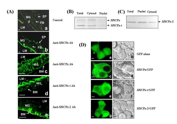
Figure 6.

Localization of SlSCPx, SlSCPx-t and SlSCPx-2 proteins. (A) Immunohistochemistry localization of SlSCPx, SlSCPx-t and SlSCPx-2 proteins in the 2-day-old 6th instar larvae of S. litura; (B and C) subcellular localization of the proteins in epithelial cells of the midgutand in the transfected Spli-221 cells (D). Anti-SlSCPx, SlSCPx-t and SlSCPx-2 antibodies were used at a dilution of 1:200 in (A). The proteins for western blotting analysis were extracted from the cytoplasm and nuclei of the midgut epithelial cells of 3-day-old old 6th instar larvae. Following electrophoresis of 20 μg protein/lane blots were probed by anti-SlSCPx-t (B) and anti-SlSCPx-2 (C) antibodies, respectively. The bands in (B) run at approximately 58 kDa and 44 kDa, respectively, and the band in (C) at approximately 16 kDa. (D) Spli-221 cells were transfected with pEGFP1 (Da and Db), pESlSCPx/GFP (Dc and Dd), pESlSCPx-t/GFP (De and Df) and pESlSCPx-C/GFP (Dg and Dh) plasmid DNAs, respectively. The photographs were taken through fluorescence (Da, Dc, De and Dg) and visible light filters (Db, Dd, Df and Dh) at 24 h post transfection. MG: midgut; EP: epidermis; FB: fat body; LM: lumen; BM: basal membrane. The scale bars represent 60 μm in (A) and 10 μm in (D).
