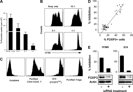
Fig. 2.
The inhibitory function of MBP83–99-specific T cells and their correlation with FOXP3. (A) CD4+CD25− T cells (responder cell or Resp.) were stimulated with anti-CD3 and anti-CD28 antibodies. FOXP3high T cells (regulator cell, clone 2C6) were added to the culture at different responder to regulator ratio. After 72 h, cells were pulsed with [3H]TdR and harvested for c.p.m. determination. (B) In another parallel experiment, responder cells were labeled with CFSE and the proliferation was measured by CFSE signal by flow cytometry. (C) The proliferative ability of FOXP3high cells was analyzed by CFSE dilution. CFSE-labeled cells were stimulated with anti-CD3 and anti-CD28 antibodies for 72 h. CFSE dilution was determined by flow cytometry. Purified CD4+CD25− T cell and CD4+CD25+ Treg were used as controls. (D) Correlation of FOXP3 expression with suppressive function was analyzed in a total of 42 MBP83–99-specific T-cell clones. Suppressive function was assessed by the inhibition of the proliferation of autologous CD4+CD25− T cells activated by anti-CD3 and anti-CD28 antibodies. Clones with an inhibition rate >50% were considered Tregs for this study. (E) Inhibitions of clones 1F5#9 and 2C6 after the treatment with siRNA specific for FOXP3 were examined by the proliferation of activated CD4+CD25− T cells. Effectiveness of FOXP3 repression was assessed by western blot for samples transfected with negative control siRNA (Ambion) or FOXP3 siRNA.

