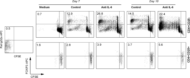
Fig. 5.
Expansion of antigen-induced FOXP3high Tregs from naive CD4+CD25− T-cell pool. PBMCs were obtained from MS patients that had significant response to MBP83–99 peptide stimulation as evidenced by proliferation assay. Purified CD4+CD25− T cells or CD4+CD25+ Treg cells (Miltenyi) from PBMC were pre-treated with 5 μM CFSE and cultured with 10 μg ml−1 MBP83–99 peptide plus irradiated APC in the presence of anti-IL-6 neutralizing antibody or isotype-matched control antibody. At day 7 and 10 after initial stimulation, cells were harvested and stained with anti-FOXP3 antibody. Cell division was analyzed by flow cytometry. The upper left quadrant is used to determine the frequency of FOXP3 up-regulated cells in CD4+ T cells.

