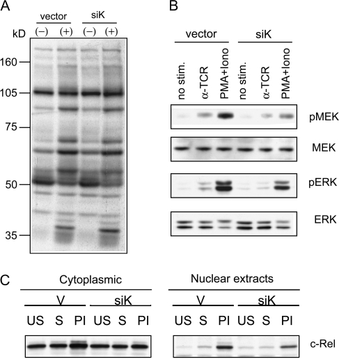
Fig. 5.
Impaired ERK activation by TCR in siK-treated Jurkat cells. Jurkat cells were transfected with siRNA expression construct for hnRNP-K (siK) or control vector (vector). (A) Transfected cells were stimulated (indicated with + above lanes) with 1 μg ml−1 soluble anti-TCR antibody or left unstimulated (−) for 5 min. Cell lysates were prepared and immunoblotted with anti-phospho-tyrosine antibody. (B) Transfected cells were stimulated with anti-TCR antibody (αTCR), PMA + iono or left unstimulated (no stim.) for 5 min. Same amount of cell lysates from each sample were analyzed by western blot with antibodies shown on the side of each panel. (C) siK- or vector-transfected cells were left unstimulated (US), stimulated with anti-TCR antibody (S) or with PMA plus ionomycin (PI). After 24 h, cells were lysed and cytoplasm and nuclear extracts were prepared for western blot analysis with anti-c-Rel antibody.

