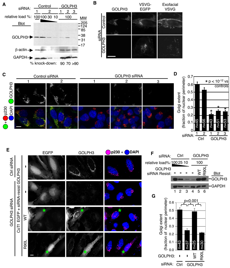
Figure 3. GOLPH3 is required for trafficking and extended ribbon morphology of the Golgi.
(A) Western blot of HeLa lysates shows GOLPH3 knocked down 70–90% by each specific siRNA. GOLPH3 antiserum recognizes a single band at 34 kDa in control lysates. Decreasing amounts of control lysate loaded to allow quantification. Blots for β-actin and glyceraldehyde-3-phosphate dehydrogenase (GAPDH) verify equal loading. (B) Trafficking of ts045-VSVG-EGFP, through Golgi to PM impaired by knockdown of GOLPH3. HeLa cells serially transfected with GOLPH3 or control siRNA, then ts045-VSVG-EGFP expression vector, incubated at 40°C overnight, then shifted to 32°C for 2 hours. Arrival at PM determined with antibody to extracellular domain applied to unpermeabilized cells (exofacial VSVG). (C) Knockdown of GOLPH3 causes condensation of Golgi ribbon. Control siRNA transfected cells show normal extended Golgi ribbon detected by GOLPH3 (green) and p230 (red). DAPI in blue. GOLPH3 siRNA reduces GOLPH3 and causes condensation of Golgi (p230). Imaging parameters identical in all. (D) Quantification of Golgi extent (Golgi length relative to nuclear perimeter, mean/SEM graphed) demonstrates significant change (p<10−12, t-test). (E) Expression of siRNA-resistant GOLPH3 (expressed near endogenous level, cotransfected with EGFP) rescues Golgi ribbon, validating GOLPH3 siRNA specificity. Expression of siRNA-resistant GOLPH3 PtdIns(4)P binding pocket mutant does not rescue Golgi ribbon phenotype. Asterisks indicate transfected cells. (F) Western blot shows knockdown of GOLPH3 (lane 4) and restoration by siRNA-resistant wild type (lane 5) and R90L (lane6) GOLPH3. Blot for GAPDH shows equal loading. (G) Quantification of Golgi extent (mean/SEM) demonstrates significant rescue by wild type, but not R90L GOLPH3 (p<0.001 for indicated comparisons, t-test).

