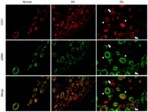
Figure 1. Detection of immature or mature blood vessels in RA synovial tissues.
Double immunoflurescent labeling of endothelium (CD31, red fluorescence) and pericytes/smooth muscle cells (aSMA, green fluorescence) in normal and RA synovial tissue is shown. Original magnification ×400. Right panels show the same area as in middle panels with higher magnification. Mature CD31+ vessels covered by aSMA+ periendothelial cells are marked by arrows, and immature CD31+ vessels lacking aSMA+ mural cells by arrow heads.

