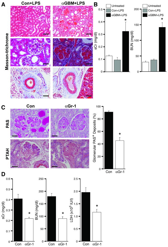
Figure 1. Development of a murine thrombotic glomerulonephritis (TGN) model.
(A) Kidneys were harvested from mice injected with control rabbit serum with LPS or two intravenous injections of anti-GBM serum (αGBM) at 1 hr intervals followed by LPS. Representative Masson-trichrome stained sections of kidneys harvested 96hr after induction of TGN are shown. Only mice given αGBM antibody plus LPS exhibited massive glomerular thrombosis, tubular dilatation and casts (top and middle panels), and plugging of large vessels (bottom panel). Bar: 100 μm (top, bottom), 30 μm (middle). (B) Serum creatinine (sCr) and blood urea nitrogen (BUN) levels were evaluated in WT mice before (Untreated) and 96 hrs following injection with control (Con) or αGBM serum plus LPS. *P<0.05 compared with Con. (C) Mice treated with control IgG (Con) or neutrophil immunodepleting anti-Gr-1 (αGr-1) were subjected to TGN. Shown are representative PAS- and PTAH- stained sections that demonstrate a partial reduction in glomerular thrombus formation (PAS), and fibrin deposition (PTAH) in αGr-1 treated mice. Scoring of PAS positive glomeruli is presented. Bar: 30 μm (D) Neutrophil immunodepletion partially attenuated serum Cr (sCr) and BUN, biochemical markers for renal function, and LDH, a general marker for organ damage. Data represent the mean ± SEM. *P<0.05 compared with Con.

