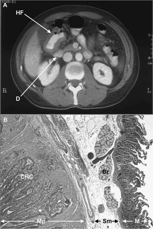
Figure 1.
(A) Computed tomography scan of a 52-year-old patient with a hepatic flexure (HF) colon adenocarcinoma adjacent to the duodenum (D). The patient underwent right colectomy and en bloc partial duodenectomy. (B) A photomicrograph of the lesion (H&E stain, ×20) demonstrating moderately differentiated colonic adenocarcinoma (CRC) invading into the muscularis propria (Mp) of the duodenum. Sm, small bowel submucosa; Br, Brunner’s glands; M, mucosa.

