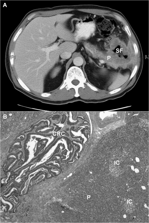
Figure 2.
(A) Computed tomography scan of a 62-year-old patient with a splenic flexure (SF) colon adenocarcinoma invading the tail of the pancreas (P) and into the abdominal wall. The patient underwent left hemicolectomy with en bloc resection of the pancreatic tail, spleen, abdominal wall, and portion of the left hemidiaphragm. (B) A photomicrograph of the lesion (H&E stain, ×20) revealing moderately differentiated colonic adenocarcinoma (CRC) invading into the pancreas (P). IC, Islet cells of Langerhans surrounded by pancreatic acinar cells.

