
Figure 4.
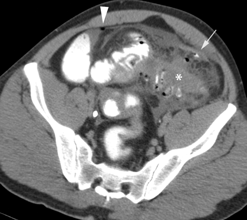
Severe diverticulitis with perforation. Computed tomography (CT) image of a 44-year-old man presenting with abdominal pain, fever, and rebound tenderness. Axial image shows sigmoid wall thickening (asterisk) and extensive fat stranding as well as small amounts of free fluid, extraluminal air (arrowhead), and trace extravasated luminal contrast (arrow).