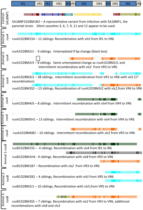
Figure 4. Low complexity of vlsE recombination events in ruvA and ruvB mutants.
The positions of the six variable regions (VR1–VR6) and six relatively invariant regions (IR1–IR6) within the vlsE central cassette region are provided at the top of the figure. The locations and silent cassette sources of the most likely recombination events for each clone depicted are indicated. A representative clone from a 28 day infection with the parental strain 5A18NP1 is shown at the top. The remainder of the figure shows the vlsE variant clonotypes isolated from C3H/HeN mice 28 days post inoculation with the ruvA mutant T11P01A01 or the ruvB mutant T03TC051. The possible involvement of each of the silent cassettes in sequence variation was analyzed using an Excel®/Visual Basic program, as described previously [11] and depicted in Fig. S2. The horizontal colored bars represent regions of each silent cassette (vls2 to vls16, top to bottom) that match the sequence changes found in the variant clone. Dark regions in each bar correspond to the actual sequence changes, whereas the lighter portion of the bar represents the maximum possible region of that silent cassette exchanged into vlsE to produce the observed sequence change. In isolates from Animal 2 inoculated with the ruvA mutant, a 9 bp untemplated change (black hatched box) that did not match the vls silent cassettes or any other genomic sequence was present in all clones isolated.

