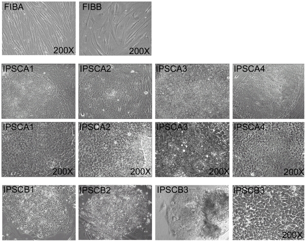
Figure 2. Phase-contrast images of FIB and IPSC lines used in this report (as labeled).
FIBA and FIBB displayed a flat stellate cytoplasm with irregular edges characteristic of fibroblast cell types (200X). IPSC lines displayed round colonies with regular edges evident in low magnification 40X images that were composed of tightly packed cells with prominent nucleoli (200X, lower panels of IPSC lines) that appeared morphologically homogenous within the center of the colony and flattened toward the edges where they were bounded by the MEF feeder layer. IPSCB1 and IPSB2 are shown only at 40X magnification.

