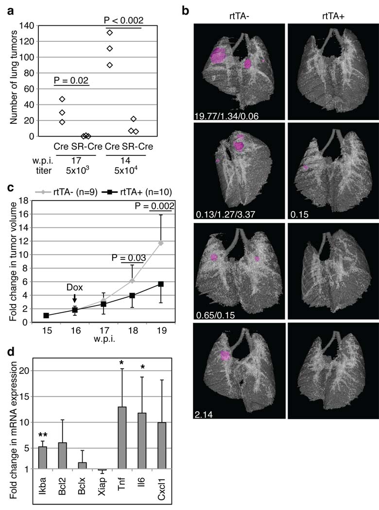
Figure 4.
NF-κB inhibition impairs lung adenocarcinoma development. (a) K-rasLSL-G12D/WT; p53Flox/Flox mice were infected with Lenti-Pgk.Cre or Lenti-UbC.IκB-SR; Pgk.Cre viruses, sacrificed 17 (left) or 14 (right) weeks post-infection (w.p.i.) and tumor number was counted (see Methods). (b–d) K-rasLSL-G12D/WT; p53Flox/Flox; CCSP-rtTA+ (rtTA+) or -rtTA− mice (rtTA−) were infected with Lenti-TRE.IκB-SR; Pgk.Cre viruses (104 viral particles). (b) From the day of infection, mice were fed a doxycycline diet (Dox). At 15 w.p.i., µ-CT imaged tumors (magenta) and lungs were reconstructed and individual tumor volumes (mm3, white text) were measured. (c) From 15 to 19 w.p.i., lungs were µ-CT imaged, and individual tumor volumes were measured. Data points represent means of fold change +/− s.d. in tumor volume relative to 15 w.p.i. (set to 1). (d) After one week of Dox treatment, RNA was isolated from tumors of rtTA+ (n=4) or rtTA− (n=3) mice. cDNA was amplified using probes for the indicated genes or Gapdh (internal control). Data show means +/− s.d. **, P=0.001; *, P=0.04 (Tnf) or 0.05 (Il6).

