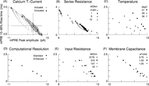
Fig. 4.
Control trials: currents and parameters. Black squares indicate baseline settings in this report. (A) Inclusion of 4.7 nA of the low threshold Ca2+ T-current led to mPREs that were slightly smaller than when excluded. All active currents, except for those carried by Ca2+, had been blocked during experiments. (B) Higher settings for the voltage clamp's series resistance led to more precise (low scatter), but less accurate (offset) recordings of synaptic currents. (C) Recording chamber temperature influenced variability through Ipre kinetics, with cooler temperatures being both more precise and more accurate. (D) Doubling the number of compartments and halving the integration time step led to minute changes, indicating that mPRE scatter (Fig. 2C) is a signature of the dendritic arbor. (E) Drastic changes in somatic Rin led to small changes in amplitudes, suggesting that recording solutions are free to be chosen based on functions other than rendering the neuron more “compact”. (F) Specific membrane capacitance influenced amplitude variability and temporal variability to roughly equal extents. (A–F) Note the preserved spatial relations (approximate co-linearity and relative inter-point distances) between the data of the five synapses.

