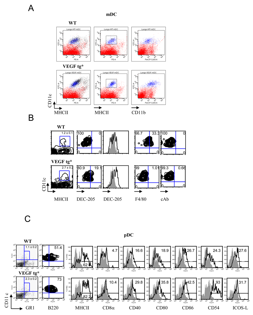
Figure 2.
Lung VEGF expression increases the number and activation of myeloid (A, B) and plasmacytoid (C) DC in tg mouse lung. Mouse lung tissues were obtained on day 7 of DOX water administration and processed as described in Materials and Methods. Single cell suspensions were stained with corresponding Ab and analyzed by flow cytometry. The results shown represent individual mouse in one out of three experiments. (A–B) Autofluorescent macrophages (shown in black color in panel A) were removed from the DC analysis. An upregulation of MHCII, CD11b, DEC-205 but not F4/80 expression on tg lung mDC was detected. For histograms: solid line represents isotype control rat IgG2a staining whereas transparent line shows the level of DEC-205 expression. *p<0.0025, WT vs tg lung mDC number (n=5/group). (C) Mouse lung pDC number increases in VEGF tg mice as compared to WT counterparts (1.1 ± 0.2 vs 2.3 ± 0.2, correspondingly, n=6, p<0045). Autofluorescent macrophages and mDC (CD11chigh) were gated out of this analysis. Histograms demonstrate upregulation of selected markers on lung pDC in VEGF tg mice.

