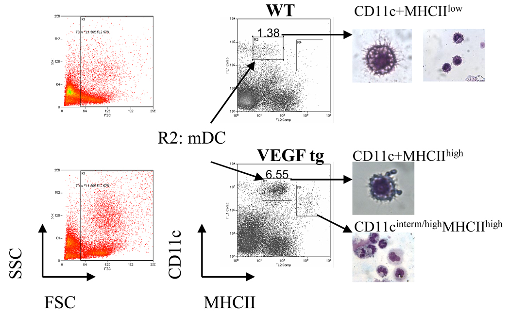
Figure 3.
Sorted lung mDC acquisition and morphology. Single cell suspensions from WT and VEGF tg mouse lungs (n=4–5 mice per experiment) prepared with omitting enzymatic digestion step were stained with a-CD11c and a-MHCII Ab, analyzed using either CellQuest, FACSDiva, or Summit software on cells sorters, and then assigned cell populations were sorted and analyzed morphologically as described in Materials and Methods. The sorted mDC selected for further characterization represent CD11c+MHCIIlow population of cells in WT mice and CD11c+MHCIIhigh population in VEGF tg mice (gate R2). CD11cinterm/highMHCIIhigh cells in gate R4 displayed a granulocyte-like morphology.

