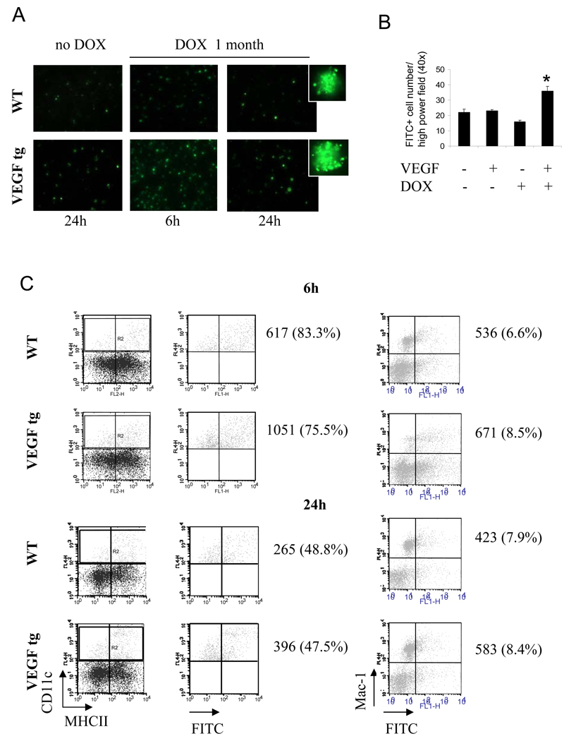
Figure 7.
VEGF-induced increase of in vivo Ag uptake by lung DC but not by Mac-1+ macrophages (A–C). WT and VEGF tg mice were kept on either normal or DOX-containing water. OVA-FITC was applied i.n. as described in Materials and Method. Single cell suspensions from lung tissues were analyzed for FITC-positivity by either fluorescent microscopy of cytospinned cells (A–B) or flow cytometry (C), n=2 mice/group/experiment in 3 separate experiments. (A) Fluorescent photomicrographs taken from WT and VEGF tg lung cells at 6h and 24h after intranasal OVA-FITC application. (B) *p<0.004, tg vs WT mice on DOX, p<0.022, tg mice on DOX vs WT and tg without DOX. (C) Lung DC (gate R2) were re-gated on OVA-FITC+. Shown is the percentage of OVA-FITC+ cells among the gated lung DC (left panel) and the percentage of CD11c-Mac-1+OVA-FITC+ cells (right panel).

