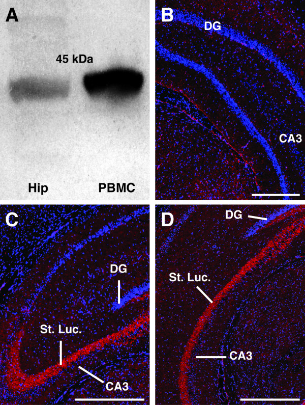
Figure 1.
CD88 expression within the hippocampal stratum lucidum. (A) Western blot demonstrating the presence of CD88 protein within the rat hippocampus (Hip). A single band was observed at 45 kDa. A similar immuno-reactive band was found in peripheral blood mononuclear cells (PBMC) that are known to express CD88. (B) Control section immunolabelled without mouse anti-ratCD88 antibody. No labelling was present within the stratum lucidum in these sections. (C and D) Intense immunolabelling for CD88 was observed within the stratum lucidum (St. Luc.) of the hippocampus (red). This immunolabelling was seen throughout the rostrocaudal extent of the hippocampus: (C) rostral hippocampus, (D) caudal hippocampus. Blue labelling in all sections is DAPI-stained nuclei. CA3, cornu ammonis 3; DG, dentate gyrus; St. Luc., stratum lucidum. Scale bar = 200 μm.

