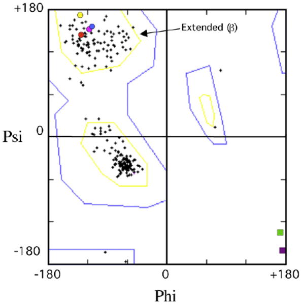
Fig. 9.

Ramachandran plot showing ϕ/φ-angles for all valines, leucines and cysteines (crosses) from X-ray structures of Dpo4 [11] hDNAP κ [18] and scDNAP κ [15], along with the flue-handles for scDNAP η (blue circle, V54), hDNAP η (red circle, V37), UmuC(V) (yellow circle, L30) Dpo4 (pink circle, C31), which are all clustered in the region expected for extended (β) secondary structure. The glycine flue-handles for hDNAP κ (G131, green square) and DNAP IV (G32, purple square) lie outside this clustered region.
