Figure 1.

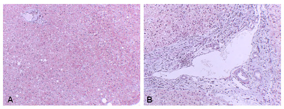
Liver fibrosis. The liver section stained with Masson's trichrome from a control (A), compared with a BDI patient (B) showed significant deposition of ECM proteins (blue stain) in B.

Liver fibrosis. The liver section stained with Masson's trichrome from a control (A), compared with a BDI patient (B) showed significant deposition of ECM proteins (blue stain) in B.