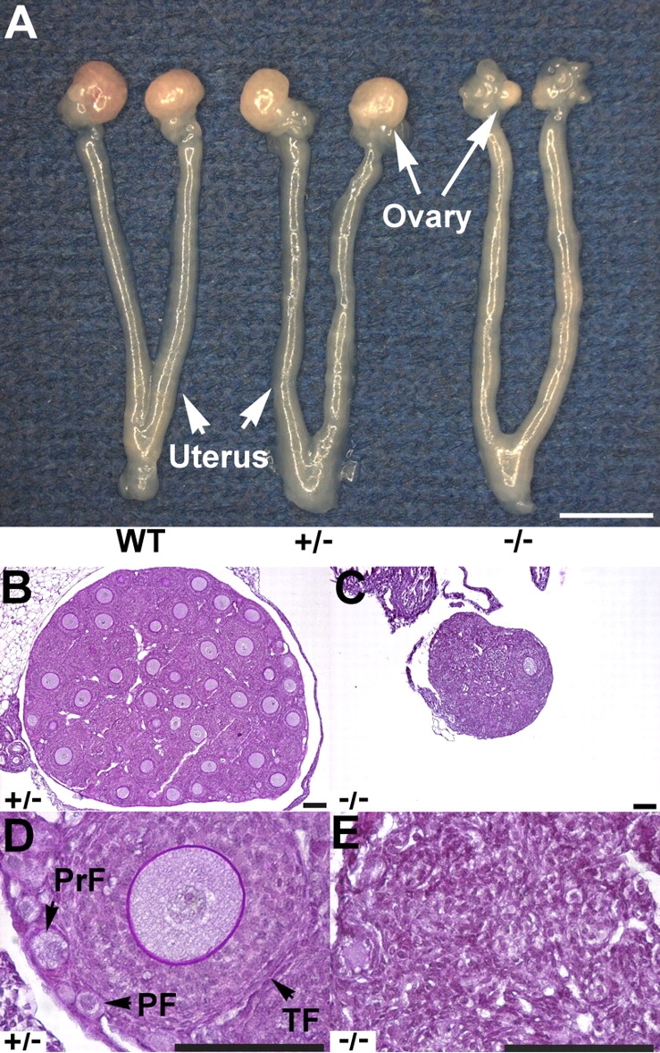
FIG. 2.

Sohlh2-knockout anatomy and histology. A) Female reproductive tracts were dissected from 3-wk-old WT, Sohlh2+/–, and Sohlh2–/– mice. Note atrophic ovaries and uterine horns in the Sohlh2–/– mice. Bar = 3 mm. B–E) Histology of 3-wk-old Sohlh2+/–and Sohlh2–/– ovaries. Sohlh2–/– ovaries lack follicles, while a magnified view of the Sohlh2+/– ovary shows well-defined primordial follicle (PF), primary follicle (PrF), and growing follicle (TF). At least five pairs of ovaries were examined, and representative sections are shown. Bar = 100 μm.
