
FIG. 2.
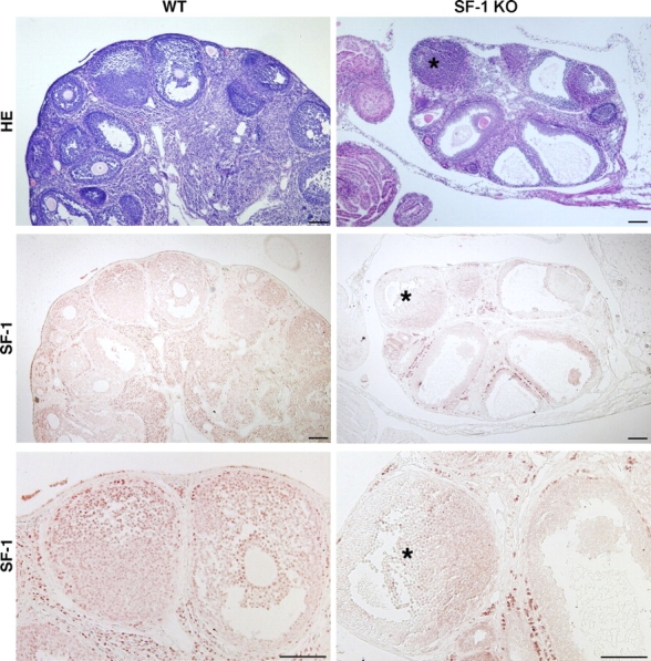
Hematoxylin-eosin staining of ovarian histology of adult WT and granulosa cell-specific SF-1 KO mice reveals the decreased size of the KO ovary and the absence of CLs. For SF-1 immunoreactivity, lower-magnification (middle panels) and higher-magnification (bottom panels) views are shown of sections of WT and granulosa cell-specific SF-1 KO mice that were analyzed for SF-1 expression by immunohistochemistry as described in Materials and Methods. The asterisk indicates a follicle in the KO section in which granulosa cells still retain some SF-1 immunoreactivity. Bar = 200 μm.