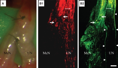
Fig. 1.
ESN of McN to UN. (A) Low-power photograph of the nerves following ESN under a stereoscope during surgery. Note that the suture was on superficial structures of the nerve. B1 and B2 are a pair of montages of low-power fluorescence micrographs of a longitudinal section of the neurorrhaphied McN and UN. UN axons were labeled by applying retrograde tracer DiI (B1, red fluorescence) from its far distal part at the time of neurorrhaphy and nerve fibers that grew into the recipient nerve were labeled retrogradely with FB (yellowish green fluorescence) from the recipient nerve 4 months following ESN. Note the continuation of yellowish-green fluorescence structures from proximal UN to the coaptated McN (B2), demonstrating the growth of nerve fibers from the presumably intact donor nerve into the recipient. The pair of fine arrows in B1 and B2 point to a cross-sectioned blood vessel, thick arrows point to the suture and arrowheads in B2 outline the epineurium. Bar = 360 μm for A and 130 μm for B1 and B2.

