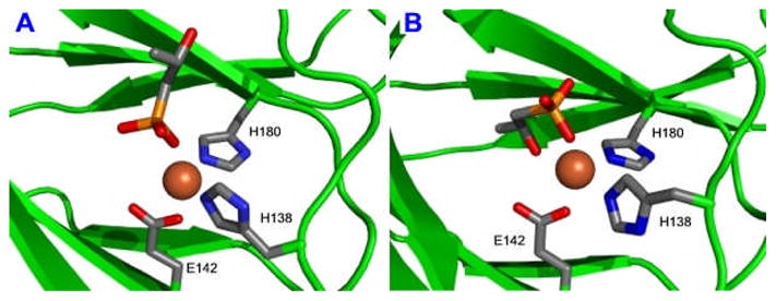
Figure 1.

X-ray Structures of the modeled HppE active site bound with Fe(II) and 2-S in monodentate (1A) and bidentate (1B) binding modes. Models were generated on the basis of the published crystal coordinates for the S. wendomorensis enzyme (PDB entries 1ZZ7 and 1ZZ8).
