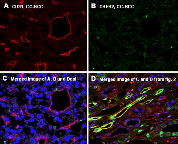
Fig. 4.

Double staining and merged presentation of CD31 and CRFR2 in CC-RCC has been shown in a–c. CD31 specific signals in microvessels of CC-RCC (a). No positivity for CRFR2 could be observed in the vasculature of CC-RCC (b). c shows the merged image of a and b and DAPI. Merged image of c and d, and DAPI with expression of CRFR2 in the vasculature of normal kidney (d)
