Fig. 3.

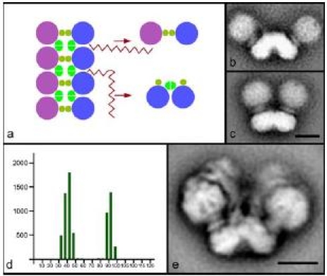
Dimers and multimers of ATP synthase. a Scheme of the multimeric chain of ATP synthases in mitochondria, which can be disrupted by detergent in two ways giving small and large angle dimers. Purple and blue circles symbolize monomeric ATP synthase complexes and ochre and bright green represent dimer-specific subunits. In mitochondria of S. cerevisiae the large angle dimers of 90° (b) and small angle dimers of 35° (c) were found in similar proportions. d Distribution of angles between the monomers of dimeric ATP synthase from S. cerevisiae. y-axis: number of particles; x-axis: size of the angle in degrees. e Dominant projection map of dimeric ATP synthase from Polytomella which corresponds to a stable dimer in which the monomers exclusively make an angle of 70°. The bars are 10 nm
