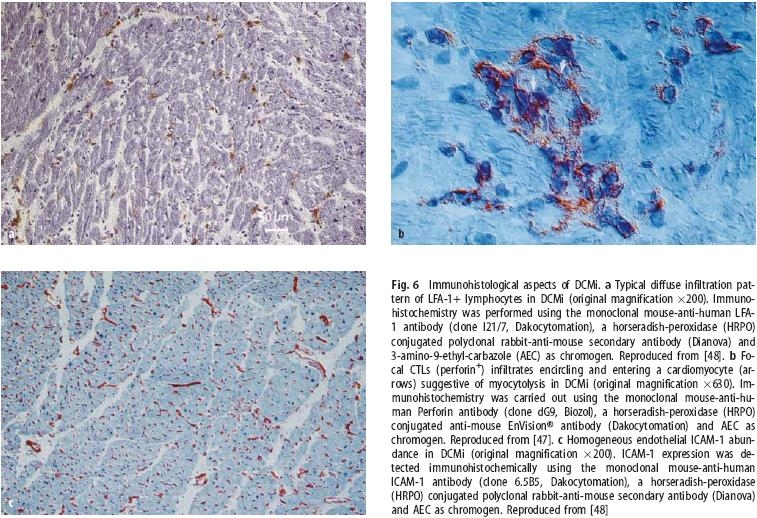
Fig. 6.

Immunohistological aspects of DCMi. a Typical diffuse infiltration pattern of LFA-1+ lymphocytes in DCMi (original magnification ×200). Immuno-histochemistry was performed using the monoclonal mouse-anti-human LFA-1 antibody (clone I21/7, Dakocytomation), a horseradish-peroxidase (HRPO) conjugated polyclonal rabbit-anti-mouse secondary antibody (Dianova) and 3-amino-9-ethyl-carbazole (AEC) as chromogen. Reproduced from [48]. b Focal CTLs (perforin+) infiltrates encircling and entering a cardiomyocyte (arrows) suggestive of myocytolysis in DCMi (original magnification ×630). Immunohistochemistry was carried out using the monoclonal mouse-anti-human Perforin antibody (clone dG9, Biozol), a horseradish-peroxidase (HRPO) conjugated anti-mouse EnVision® antibody (Dakocytomation) and AEC as chromogen. Reproduced from [47]. c Homogeneous endothelial ICAM-1 abundance in DCMi (original magnification ×200). ICAM-1 expression was detected immunohistochemically using the monoclonal mouse-anti-human ICAM-1 antibody (clone 6.5B5, Dakocytomation), a horseradish-peroxidase (HRPO) conjugated polyclonal rabbit-anti-mouse secondary antibody (Dianova) and AEC as chromogen. Reproduced from [48]
