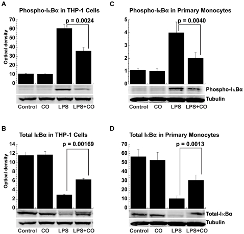
Figure 8. CO prevention of LPS-induced IκBα phosphorylation and degradation.
(A) Phosphorylated IκBα in THP-1 cells, (B) total IκBα in THP-1 cells, (C) phosphorylated IκBα in primary human monocytes, and (D) Total IκBα in primary human monocytes. Whole cell extracts or cytoplasmic fractions were prepared from THP-1 cells (2×10⁶) and elutriated primary human monocytes (5×10⁶), treated with or without LPS (1 µg/ml) in the presence or absence of CO (250 ppm) for 15 min and examined for phosphorylated or total IκBα by Western blotting. α-Tubulin served as a equal loading and transfer control. Densitometry results are shown as the means ± SEM of three independent experiments.

