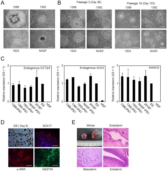
Figure 2. Generation and maintenance of human iPS cells on autologous feeders.
A. Images of primary iPS cell colonies. We introduced 4 reprogramming factors into 1388, 1392, 1503 or NHDF. The colonies were photographed 25 days after transduction. Bars indicate 200 µm. B. Images of established iPS clones. We isolated iPS clones and transferred onto each isogenic fibroblast line. The cells at passage 3 and 18 were photographed. Bars indicate 200 µm. C. Quantification of the expression of pluripotent stem cell markers. Total RNA of iPS cells established from four independent fibroblast lines maintained on isogenic feeders, H9 ES cells and HDF was purified, and used for reverse transcription. The graphs show the average of three independent experiments. Error bars indicate standard deviation. Data were normalized with the results of G3PDH. D. In vitro differentiation of iPS cells. iPS cells were transferred to suspension culture to form embryoid bodies for 8 days. Embryoid bodies were transferred to gelatin-coated plated, and incubated another 8 days. The cells were stained with anti-SOX17 (red), anti-α-SMA (red) or anti-NESTIN (green) antibodies. Nucleuses were stained with Hoechst 33342 (blue). Bars indicate 100 µm. E. Teratoma formation of iPS cells. Paraffin-embedded sections were stained with hematoxylin and eosin.

