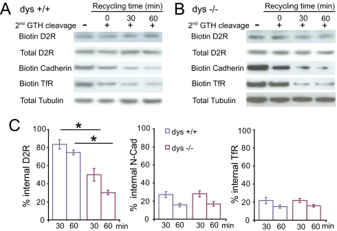
Fig. 2.
Increased Recycling of Internalized D2 Shown by Biochemical Assay. (A and B) Biotinylation assay of D2 recycling in wild-type (A) and dys−/− (B) cortical neurons. Surface proteins were labeled with cleavable biotin, treated with dopamine (10 μM) at 37 °C for 60 min (internalization), and the residual surface biotin was removed by a first round glutathione treatment. The internalized membrane proteins were then allowed to recycle back to cell surface at 37 °C for 30 or 60 min. A second round of glutathione was applied to cleave any appearing surface biotin. Representative immunoblots show remaining intracellular D2, N-Cad, and TfR. A control experiment (left lane) was performed at 4 °C to prevent recycling after endocytosis and internalized proteins remained inside the cells. The loss of biotinylated D2 after a second biotin cleavage provides a measure method of receptor recycling. (C) Quantitative analysis of D2 recycling. The remaining internalized D2 was normalized to that presented at time 0. *, P < 0.05.

