Fig. 3.
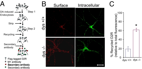
Increased Reinsertion of Internalized FLAG-D2 Shown by Imaging. (A) Schematic of recycling assay. Neurons expressing FLAG-D2 were incubated at 37 °C with M1 antibody (Ca2+ sensitive) for 20 min, followed by dopamine for 60 min. The surface M1 antibody was removed by stripping, and neurons were returned to incubator for receptor recycling. Neurons were fixed and stained with Alexa-546 conjugated secondary antibody to label surface D2 under nonpermeable conditions (red), or Alexa-488 conjugated secondary antibody (green) to label intracellular D2 under permeable conditions. (B) Representative images from recycling experiment showing recycled (Left, Surface) and intracellular (Right) FLAG-D2 after 60 min of recycling. Immunofluorescence staining of D2 was performed under non-permeable (red) and permeable (green) conditions. (Scale bar, 20 μm.) (C) Quantification of D2 reinsertion measured as the ratio of surface (red)/total (red + green) fluorescence. *, P < 0.05.

