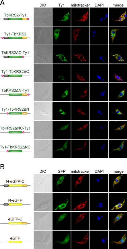
Fig. 4.
Analysis of TbKRS2 import mechanism into the mitochondria. (A) Confocal immunofluorescence analysis of different TbKRS2 forms. From left to right, cells were visualized as described for Fig. 3B. (B) Confocal immunofluorescence analysis of full-length different eGFP fusion proteins. Cells were visualized as described for Fig. 3B, but overexpressed protein (green channel) was detected directly by GFP fluorescence.

