Fig. 3.
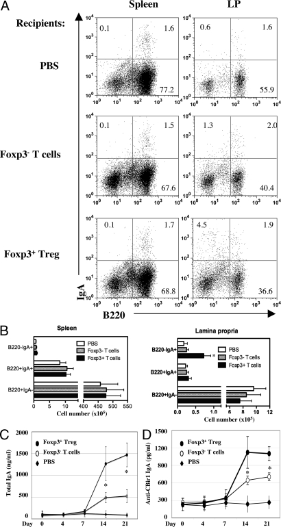
Adoptive transfer of CD4+foxp3+ Treg cells restored intestinal IgA in B6.TCRβ×δ−/− mice. CD4+ foxp3+ Treg cells and CD4+ foxp3− T cells from B6.GFP-foxp3 mice were transferred into B6.TCRβ×δ−/− mice at 1 × 10⁶/mouse. (A) LP IgA+ B cells were analyzed by flow cytometry at day 21. (B) B cell numbers from four mice of each group. *, P ≤ 0.05 compared to the recipient mice of CD4+ foxp3− T cells. (C and D) Stool pellets were collected at days 0, 4, 7, 14, and 21. Total IgA (C) and anti-CBir1 IgA (D) were assessed by ELISA. *, P ≤ 0.05 compared to foxp3− T cell group.

