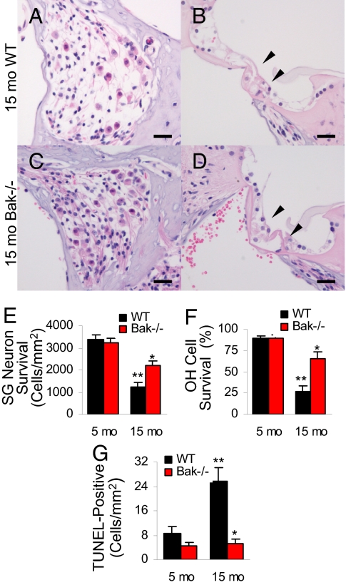
Fig. 2.
Bak deficiency reduces cochlear pathology. SG neurons (A and C) and hair cells (B and D) in the basal cochlear regions from 15-month-old WT and Bak−/− mice. Arrows indicate the hair cell regions. (Scale bar, 200 μm.) (E) SG neuron survival (i.e., SG neuron density) of basal cochlear regions was measured from WT and Bak−/− mice at 5 and 15 months of age (n = 5). (F) OH cell survival (%) of basal cochlear regions was measured from WT and Bak−/− mice at 5 and 15 months of age (n = 5). (G) TUNEL-positive cells were counted in the cochlea of WT and Bak−/− mice at 5 and 15 months of age (n = 5). **Significantly different from 15-month-old WT mice (P < 0.05), **Significantly different from 5-month-old WT mice (P < 0.05). Error bars represent SEM.

