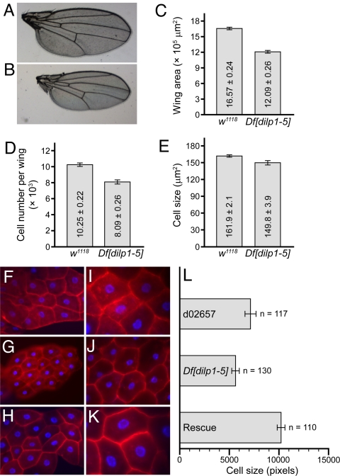
Fig. 2.
Small size of Df[dilp1–5] homozygotes is due to reduced cell size and cell number. (A–E) Cell number and cell size in dissected wings. (A and B) Photos of dissected wings from (A) adult female, w1118 control or (B) Df[dilp1–5] homozygote. (C) The overall wing area is smaller in Df[dilp1–5] homozygotes than in w1118 controls. (D and E) Measurements of cell number and cell size in w1118 or Df[dilp1–5] homozygotes. Both decreased in Df[dilp1–5] mutants. n = 5 for C–E. (F–K) Fat body cell size is reduced in Df[dilp1–5] animals. Photos of larval fat bodies from wandering third-instar larvae stained with phalloidin (red) and DAPI (blue) to reveal nuclei. [×20 (F, G, and H); ×40 (I, J, and K).] Genotypes: (F and I) Control parental line d02657, (G and J) Df[dilp1–5]/Df[dilp1–5], (H,K) hsGAL4>UASdilp2; Df[dilp1–5]/Df[dilp1–5]. (L) Quantitation of fat body cell size was carried out with ImageJ software. The cell size of Df[dilp1–5] homozygotes was approximately 80% of the parental control. Error bars indicate standard error.

